
Fig. 2
- ID
- ZDB-IMAGE-160706-8
- Antibodies
- Publication
- Raman et al., 2016 - aPKC regulates apical localization of Lgl to restrict elongation of microridges in developing zebrafish epidermis
- All Figures
- Figures for Raman et al., 2016
|
Fig. 2
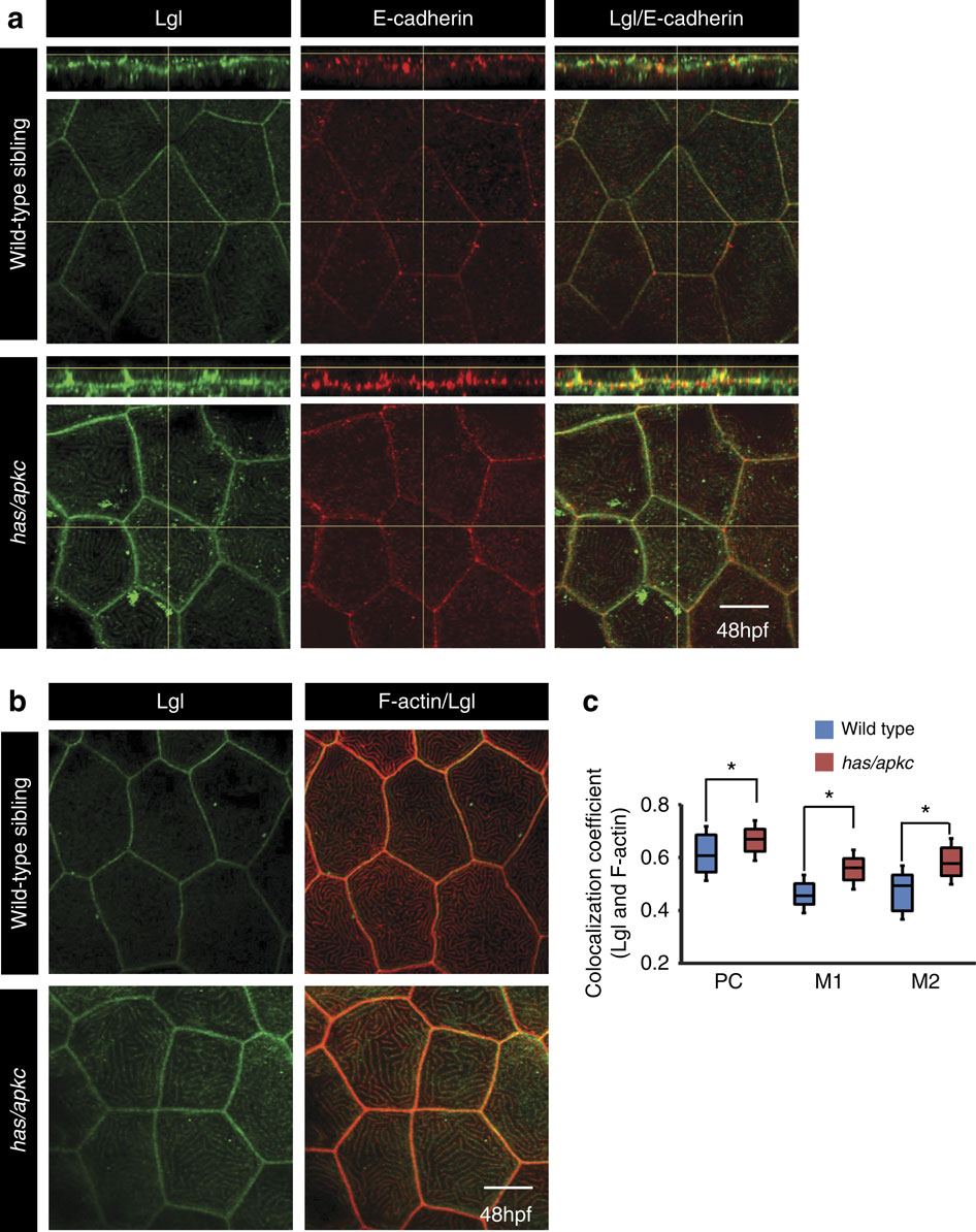
The levels of apical Lgl increase in the apical domain of the head peridermal cells in has/apkc mutants.Immunostaining using anti-Lgl2 and E-cadherin antibodies (a) in wild-type and has/apkc mutant embryos. Immuno-colocalization of Lgl and F-actin in the apical domain of wild-type and has/apkc mutant embryos at 48 hpf (b). Colocalization coefficient analysis between Lgl and F-actin by Pearson’s (PC) method and Manders’ overlap for Lgl with F-actin (M1) and F-actin with Lgl (M2) at 48 hpf in wild-type and has/apkc mutant embryos (c). Asterisks in c indicate significant difference at P<0.001 by Student’s t-test. Scale bar in a and b is equivalent to 10 µm.