
|
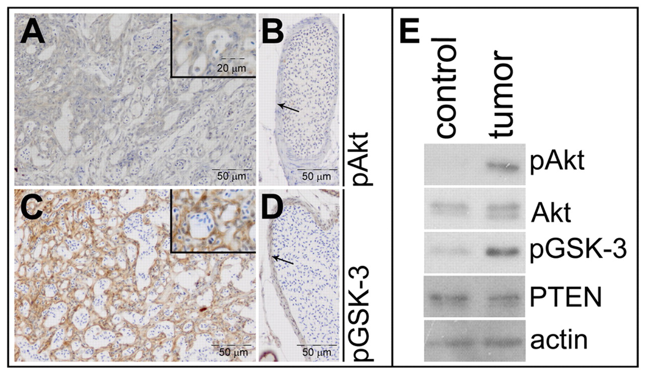
Fig. 4
Elevated Akt/PKB signaling in Pten mutant tumors. Immunohistochemistry on transversal sections from a ptena+/-ptenb-/- mutant fish, using pAkt- and pGSK-3β-specific antibodies. (A) Tumor area stains weakly positive for pAkt, whereas (B) control vessel (arrow) from the same section is not stained (n=10). (C) Tumor area is highly positive for pGSK-3β, whereas (D) cells from a control vessel (arrow) from the same section only stain mildly positive (n=6). (E) The cranial part of a ptena+/-ptenb-/- mutant that developed a tumor was dissected into two fragments, one harboring tumor tissue (tumor) and the other representing control tissue (control). The samples were lysed and the lysates were run on a denaturing SDS-polyacrylamide gel. The proteins were transferred to a PVDF membrane and after blocking the blot was probed with anti-pAkt antibody, stripped and sequentially probed with anti-Akt, anti-pGSK-3, anti-PTEN and, as a loading control, anti-actin.