
Fig. 9
- ID
- ZDB-IMAGE-160630-32
- Publication
- Lovely et al., 2016 - Bmp signaling mediates endoderm pouch morphogenesis by regulating Fgf signaling in zebrafish
- All Figures
- Figures for Lovely et al., 2016
|
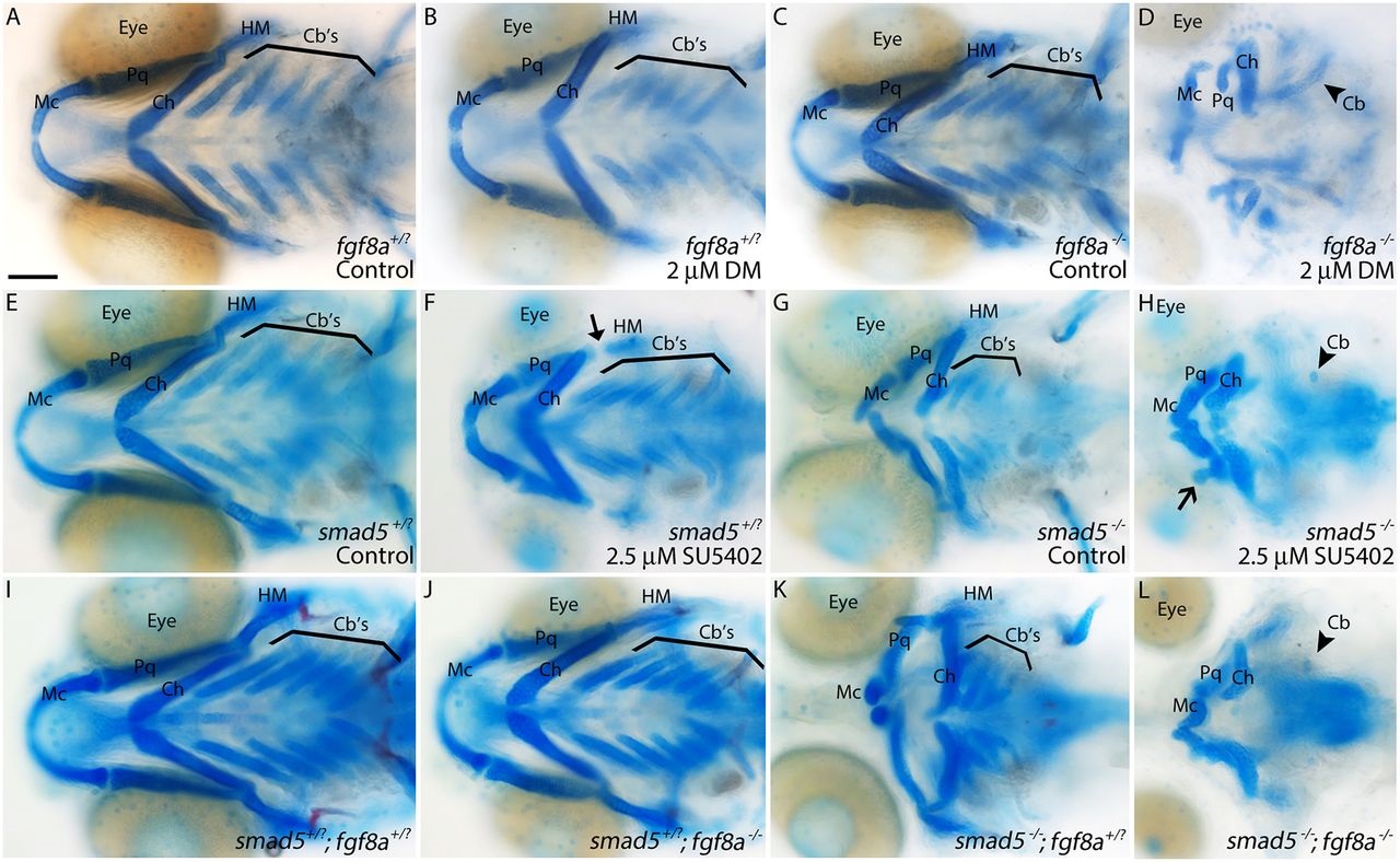
Fig. 9
Bmp and Fgf signaling synergistically interact to achieve proper craniofacial development. Whole-mount images of 5dpf viscerocranium. (A-C) No gross craniofacial defects are present in wild-type or heterozygous controls (A), wild-type embryos treated with suboptimal doses of Dorsomorphin (B) or untreated fgf8a mutants (C). (D) Treating fgf8a mutants with suboptimal doses of Dorsomorphin causes severe craniofacial defects. (E,F) Similarly, wild-type or smad5 heterozygous embryos do not display any craniofacial defects (E), whereas these embryos treated with suboptimal doses of SU5402 (Pan-Fgf inhibitor, 10-18hpf) display minor hyomandibular defects (F, arrow). (G) Untreated smad5 mutants have characterized craniofacial defects (Swartz et al., 2011). (H) Suboptimal SU5402-treated smad5 mutants display severe craniofacial defects, including loss of ceratobranchial cartilages (arrowhead). (I-L) Severe craniofacial defects mirroring those of SU5402-treated smad5 mutants (see H) are present in smad5; fgf8a compound mutants (L) and not any other allelic combination (I-K). Ventral views, anterior to the left. Cartilages: Mc, Meckel′s; Ch, ceratohyal; Cb′s, ceratobranchial; Pq, palatoquadrate; HM, hyomandibular. Scale bar: 100µm.