Image
Figure Caption
Fig. 2
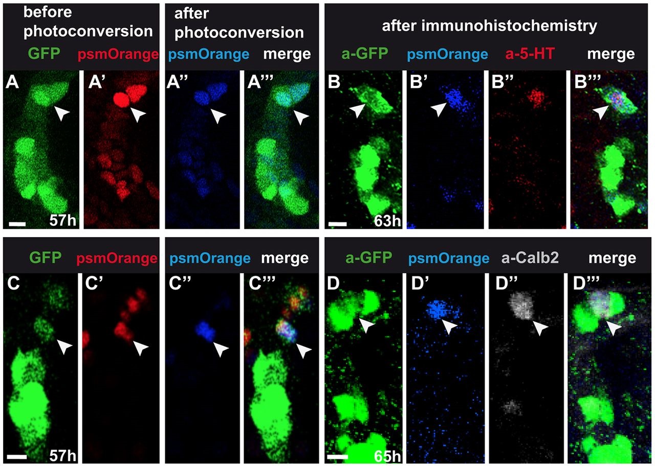
Fate-mapping of tg(fgf8a.dr17:gfp) cells. 4µm confocal projections of anterior mouth. One embryo per row, arrowheads indicate the same cell per row. (A,C) tg(fgf8a.dr17:gfp), (A′,C′) non-photo-converted, (A′′,C′′) photo-converted nuclear PSmOrange expression in forming taste buds. (B,D) tg(fgf8a.dr17:gfp) expression detected with anti-GFP antibody. (B′,D′) Photo-converted nuclear PSmOrange after fixation. (B′′,D′′) Antibody-detected 5-HT or Calb2 expression. Scale bars: 10µm.
Acknowledgments
This image is the copyrighted work of the attributed author or publisher, and
ZFIN has permission only to display this image to its users.
Additional permissions should be obtained from the applicable author or publisher of the image.
Full text @ Development

