
Fig. 5
- ID
- ZDB-IMAGE-160630-10
- Antibodies
- Publication
- Suli et al., 2016 - Innervation regulates synaptic ribbons in lateral line mechanosensory hair cells
- All Figures
- Figures for Suli et al., 2016
|
Fig. 5
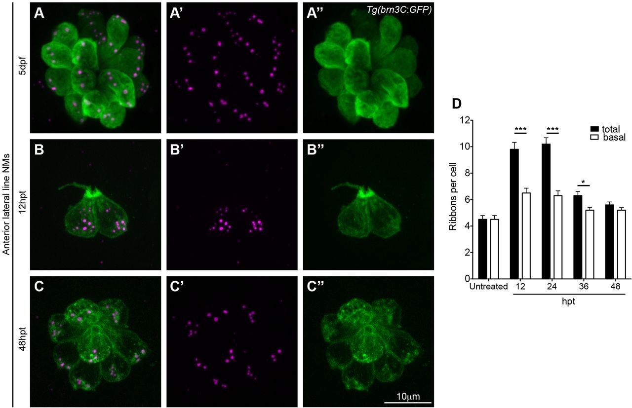
Membrane-adjacent ribbons predominate in mature hair cells. (A-C′′) 5dpf Tg(brn3C:GFP) larvae were treated with 400µM neomycin for 1h to kill all hair cells. At different time points during regeneration larvae were fixed and stained with anti-GFP antibody (green, hair cells) and anti-RibeyeB antibody (magenta, ribbons), and three anterior lateral line neuromasts (NMs) (O2, MI1, MI2, IO4 or M2) were imaged using a confocal microscope. (D) Ribbons seen right next to the basal membrane in the green channel, as visualized in 3D surface renderings (Movies 6 and 7), were counted as basal ribbons, whereas the rest of the ribbons were considered as distal ribbons. At early time points during regeneration both distal ribbons and membrane adjacent ribbons were present in hair cells (12-36 hpt). Distal ribbons eventually disappeared, and by 48h post treatment (hpt) ribbons adjacent to the basolateral membrane predominated. Ribbon numbers were obtained by visually counting ribbons in surface renderings of images of neuromasts (see Materials and Methods). Three neuromasts in three different larvae were imaged and ribbons were counted in a total of 26 hair cells of untreated larvae, 24 hair cells of 12hpt larvae, 51 hair cells of 24hpt larvae, 54 hair cells of 36hpt larvae and 65 hair cells of 48hpt larvae. *P=0.0124, ***P<0.0001 (unpaired two-tailed t-test). Results are mean±s.e.m.