Image
Figure Caption
Fig. 9
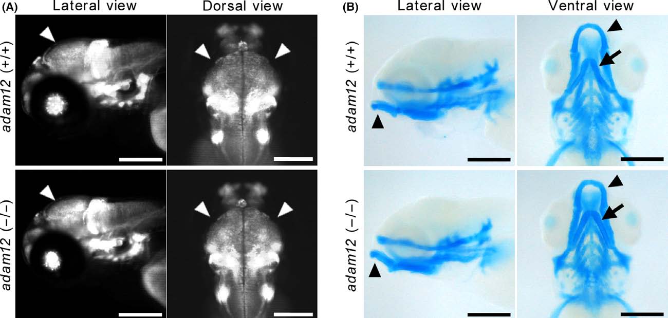
Phenotypic analysis of the cephalic nerve and jaw cartilage. (A) Cephalic nerve, including telencephalon and midbrain (arrowheads), as visualized by neurod: EGFP expression. (B) Alcian Blue staining of jaw cartilage, including Meckel′s cartilage (arrowheads) and ceratohyal (arrows) at 5-dpf. Scale bar: 200 µm.
Figure Data
Acknowledgments
This image is the copyrighted work of the attributed author or publisher, and
ZFIN has permission only to display this image to its users.
Additional permissions should be obtained from the applicable author or publisher of the image.
Full text @ Dev. Growth Diff.

