
Fig. S2
- ID
- ZDB-IMAGE-160607-8
- Publication
- Enyedi et al., 2016 - The Cell Nucleus Serves as a Mechanotransducer of Tissue Damage-Induced Inflammation
- All Figures
- Figures for Enyedi et al., 2016
|
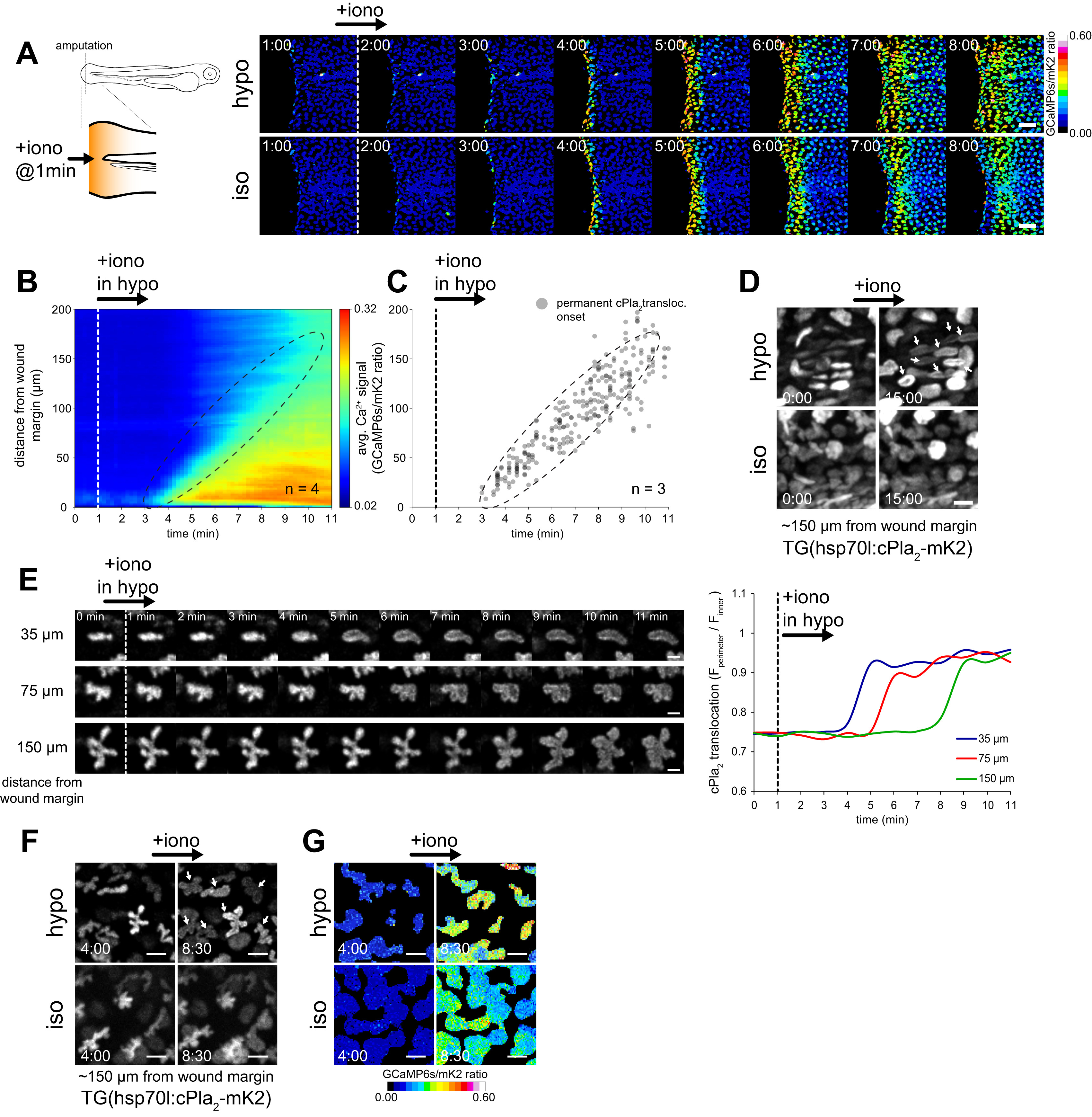
Fig. S2
Osmotic Cell Swelling and Ca2+ Signals Are Required for cPla2 Activation in Live Zebrafish, Related to Figure 1
(A) Left, scheme of experimental setup. Right, ratiometric imaging of Ca2+ transients induced by stimulation of amputated tail fins with the Ca2+-ionophore ionomycin in hypotonic (top) or isotonic (bottom) bathing solution (also see Movie S2). To allow drug penetration, tail fin tips were amputated under isotonic conditions ~10 min in advance. Scale bars, 50 µm.
(B) Average spatiotemporal Ca2+ signal profile of the indicated number of transgenic Ca2+ reporter larvae Tg(hsp70l:GCaMP6s-NLS-P2A-mK2-NLS) after ionomycin stimulation in hypotonic solution (see A, “hypo,” top).
(C) Spatiotemporal plot of permanent cPla2-mK2 INM-translocation events induced by ionomycin treatment at the indicated time under hypotonic conditions in the indicated number of Tg(hsp70l:cPla2-mK2) larvae. Dotted ellipse, corresponding regions in plots B and C.
(D) Representative confocal images showing cPla2-mK2 localization in transgenic zebrafish larvae before and after ionomycin stimulation under hypotonic (top) or isotonic (bottom) conditions, at a site ~150 µm from the wound margin (see also Movie S3). White arrows, INM-translocation of cPla2-mK2. Scale bars, 10 µm.
(E) Left panel, representative confocal images showing cPla2-mK2 localization of cells at the indicated distances from the wound margin in Tg(hsp70l:cPla2-mK2) larvae treated with ionomycin under hypotonic conditions at the indicated time. Right panel, quantification of cPla2-mK2 translocation (see Experimental Procedures for details). Scale bars, 10 µm.
(F) Confocal imaging of Tg(hsp70l:cPla2-mK2) zebrafish larvae before and after ionomycin stimulation under hypotonic (top) or isotonic (bottom) conditions, focusing on the fibroblast-like mesenchymal cells ~150 µm from the wound margin. Note the translocation of cPla2-mK2 to the nuclear envelope in the upper right panel as marked by white arrows. Scale bars, 10 µm.
(G) Confocal imaging of Ca2+ signals triggered by ionomycin under hypotonic or isotonic conditions in Tg(hsp70l:GCaMP6s-NLS-P2A-mK2-NLS) zebrafish larvae after tail fin tip amputation. A segment of the tail fin similar to the one in F is enlarged, showing fibroblast-like mesenchymal cells before and after the ionomycin-induced Ca2+ wave reaches this region. Scale bars, 10 µm.
Reprinted from Cell, 165, Enyedi, B., Jelcic, M., Niethammer, P., The Cell Nucleus Serves as a Mechanotransducer of Tissue Damage-Induced Inflammation, 1160-1170, Copyright (2016) with permission from Elsevier. Full text @ Cell