
Fig. 3
- ID
- ZDB-IMAGE-160607-5
- Publication
- Enyedi et al., 2016 - The Cell Nucleus Serves as a Mechanotransducer of Tissue Damage-Induced Inflammation
- All Figures
- Figures for Enyedi et al., 2016
|
Fig. 3
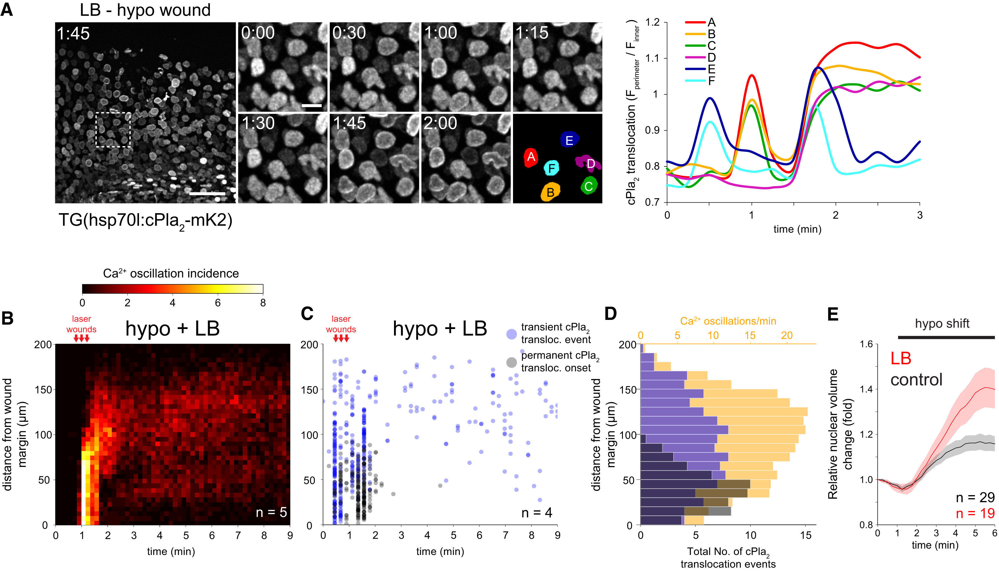
F-Actin Inhibits cPla2 Translocation and Nuclear Swelling in Live Zebrafish
(A) Left: confocal maximum intensity projections of cPla2-mK2 in latrunculin B (LB) pretreated zebrafish larvae, wounded under hypotonic conditions at t = 0.5-1 min. Note the wave of cPla2-mK2 translocation reaching ~200 µm deep into the tissue (see also Movie S4). Scale bars, 50 and 10 µm. Right: quantification of cPla2-mK2 translocation (see the Experimental Procedures for details) in selected nuclei, marked with rainbow color masks (A-F) on the left panel.
(B) Averaged spatiotemporal profile of Ca2+ signal oscillation frequency of the indicated number of transgenic Ca2+ ratio-reporter larvae, pretreated with latrunculin B (LB) and wounded at the indicated time under hypotonic conditions.
(C) Spatiotemporal plot of permanent and transient cPla2-mK2 INM-translocation events induced by wounding under hypotonic conditions after LB-pretreatment in the indicated number of Tg(hsp70l:cPla2-mK2) larvae.
(D) Average spatial distribution of cPla2-mK2 translocation events (blue, transient; gray, permanent) and Ca2+ oscillation frequency (orange) as a function of distance from the wound margin, induced by wounding LB-pretreated larvae under hypotonic conditions (data from experiments shown in B and C).
(E) Average nuclear volume evolution in the suprabasal and basal epithelial cells of zebrafish tail fin after hypotonic shifting in untreated and LB-pretreated larvae, measured by confocal imaging of nuclear targeted EGFP. Error bars, SEM; n, number of cells.
Reprinted from Cell, 165, Enyedi, B., Jelcic, M., Niethammer, P., The Cell Nucleus Serves as a Mechanotransducer of Tissue Damage-Induced Inflammation, 1160-1170, Copyright (2016) with permission from Elsevier. Full text @ Cell