
|
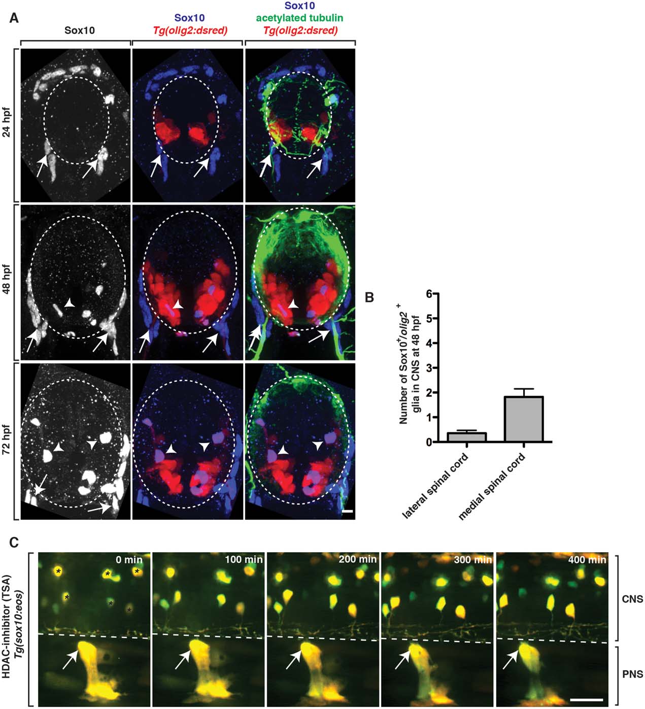
Fig. 3
OPCs do not restrict ectopic peripheral glial migration. A: Images from 24, 48, and 72 hpf Tg(olig2:dsred) embryos stained with antibodies specific to acetylated tubulin (green) and Sox10 (blue). Arrows show peripheral Sox10+ cells while arrowheads denote centrally-located Sox10+ cells. B: Quantification of the location of Sox10+ OPCs in the spinal cord at 48 hpf. C: Images from a 24 h time-lapse movie starting at 48 hpf of a Tg(sox10:eos) embryo treated with TSA at 36 hpf. Arrows denote PNS glia that do not migrate into the spinal cord. Asterisks indicate sox10+ neurons that are labeled by Tg(sox10:eos). Dashed lines and circles denote boundary of spinal cord. Scale bars, 25 µm.