
|
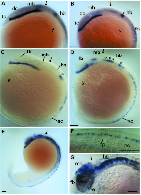
Fig. 4
Whole-mount in situ hybridization analysis of hlx-1 expression of different developmental stages. Side views (anterior to the left) of 11, 12, 13, 16, 19 and 30 hpf are shown in A, B, C, D, E and G, respectively. The spinal cord of the 19 hpf embryo is shown at high magnification in F. Arrows indicate the location of the midbrain-hindbrain boundary and open triangles mark the position of rhombomere-restricted sites of expression in the hindbrain. Bar, 60 µm. Abbreviations: dc, diencephalon; e, eye; fb, forebrain; fp, floor plate; hb, hindbrain; mb, midbrain; nc, notochord; sc, spinal cord; tc, telencephalon; y, yolk.