
Fig. S3
- ID
- ZDB-IMAGE-160524-24
- Publication
- Ohnmacht et al., 2016 - Spinal motor neurons are regenerated after mechanical lesion and genetic ablation in larval zebrafish
- All Figures
- Figures for Ohnmacht et al., 2016
|
Fig. S3
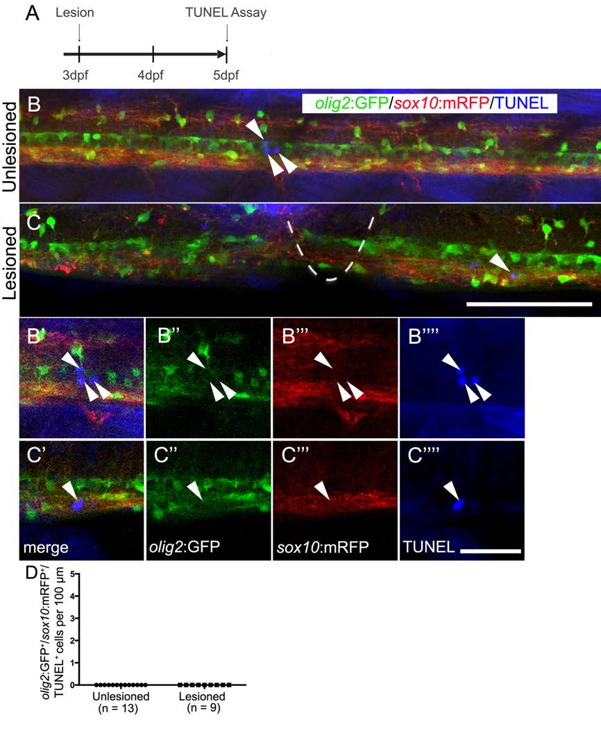
Oligodendrocyte-lineage cells do not show increased cell death after a lesion. Lateral views of the trunk area are shown; rostral is left, dorsal is up. A: The timeline of the experiment is shown. B,C: In the unlesioned and lesioned spinal cord, TUNEL+ cells (arrows) are rare (lesion site indicated by dashed line in C). B′-C′′′′: Analysis of single optical sections at higher magnification reveals lack of co-labeling of TUNEL+ cells with olig2:GFP and sox10:mRFP. D: olig2:GFP+/sox10:mRFP+/TUNEL+ cells were not observed in unlesioned and lesioned animals. Bar in C = 100 µm for B,C; in C′′′′ = 20 µm for B′-C′′′′.