
Fig. S4
- ID
- ZDB-IMAGE-160524-13
- Genes
- Publication
- Eno et al., 2016 - aura/mid1ip1L regulates the cytoskeleton at the zebrafish egg-to-embryo transition
- All Figures
- Figures for Eno et al., 2016
|
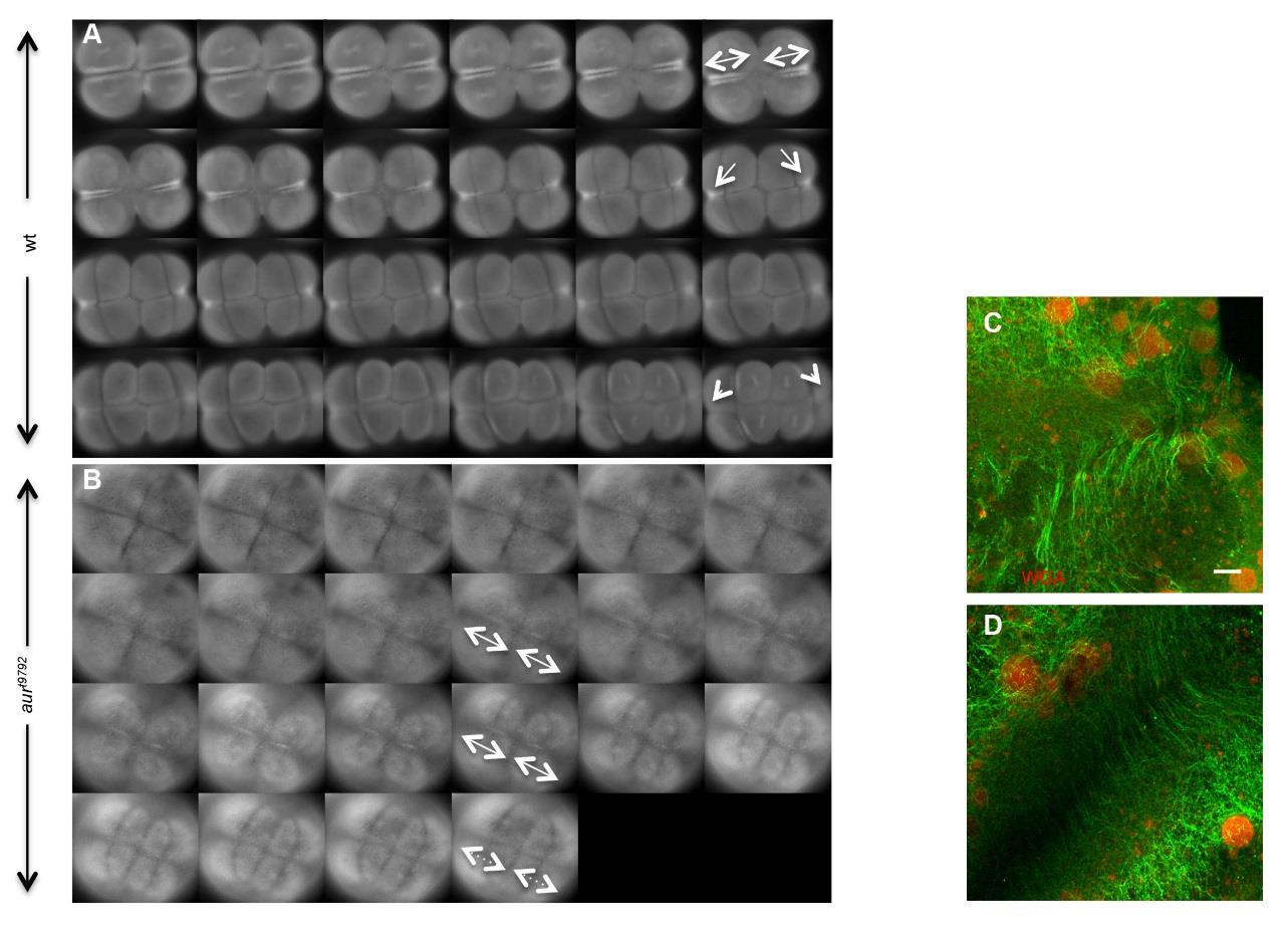
Fig. S4
Lack of FMA reorganization in aura embryos. Live imaging of microtubules in embryos using the EMTB transgene in wild-type and aura mutant line. (A) In wild-type, the FMA forms along the furrow (double arrows), compacts distally (arrows) and disappears (arrowheads). (B) In aura mutant embryos, the FMA also appears along the furrow (double arrows), but does not experience a distal shift and remains in an apparently stabilized condition until it eventually becomes undetectable (dotted line arrows). Images were taken in one minute intervals from 60 mpf to 85 mpf. (C,D) Microtubule reorganization defect occurs independently of the number of ectopic CGs in aura mutants. In aura mutants, the FMA appears stabilized in all (100%) embryos, regardless of the number of ectopic CGs at the furrow: (C) furrow with ≥ 5 GCs (6/16 examined furrows); (D) furrow with ≤ 4 CGs (10/16 examined furrows). Furrows shown are examples of the 1st furrow of a single embryo at 75 mpf, when the FMA has normally become either distally enriched or disassembled (Urven et al, 2006). Similar results are obtained with the 2nd furrow (not shown) Scale bar: C,D, 10 µm