
Fig. 1
- ID
- ZDB-IMAGE-160524-1
- Publication
- Eno et al., 2016 - aura/mid1ip1L regulates the cytoskeleton at the zebrafish egg-to-embryo transition
- All Figures
- Figures for Eno et al., 2016
|
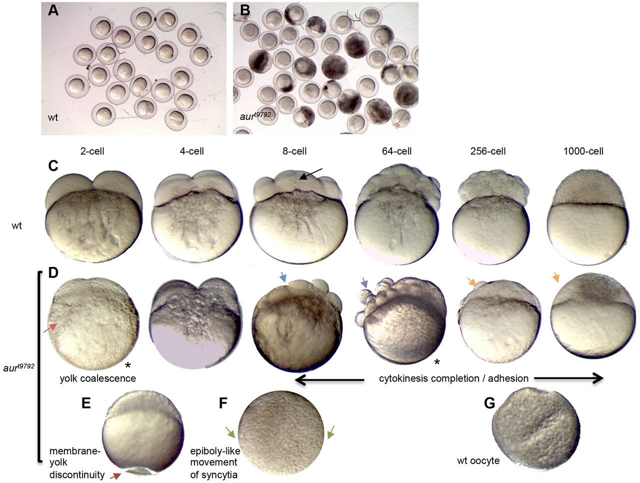
Fig. 1
Developmental timecourse of zebrafish aura mutant embryos. (A,B) Embryo clutches from wild-type (A) and aura mutant (B) mothers at 10mpf showing egg lysis in mutants. (C-G) Wild-type (C,G) and aura mutant (D-F) timecourse. aura embryos do not display normal yolk coalescence (red arrow), instead resembling inactive wild-type mature oocytes (G). At the 8-cell stage, a septum is apparent in wild type (black arrow), whereas aura mutants lack this septum (blue arrow). aura mutants subsequently exhibit rounded, non-adhesive cells (purple arrow). In the cleavage stages, aura mutant embryos are partially or fully syncytial (orange arrows). Other phenotypes include discontinuities between the egg plasma membrane and the yolk along any animal-vegetal position (E, dark red arrow). Syncytia in aura mutants undergo an epiboly-like movement (F, green arrows indicate migrating edge).