
Fig. 3 S2
- ID
- ZDB-IMAGE-160517-52
- Publication
- Dunn et al., 2016 - Brain-wide mapping of neural activity controlling zebrafish exploratory locomotion
- All Figures
- Figures for Dunn et al., 2016
|
Fig. 3 S2
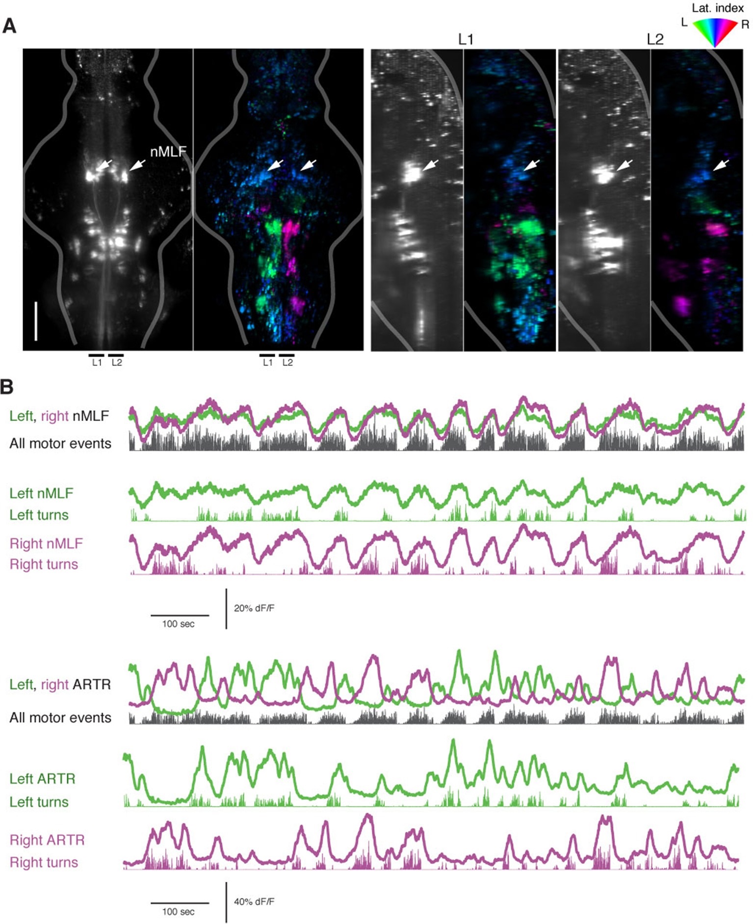
The nMLF is correlated with swim amplitude but not direction.
(A) Left, light-sheet micrograph of the backfilled reticulospinal system (grayscale, dextran-conjugated TxRed, see ′Materials and methods′) and GCaMP6f regression map from the same fish. Right, sagittal views of the same fish, projected across the regions L1 and L2, as indicated to the left. Arrows in all maps point to the overlap between zones of blue-tuned regions in the functional brain maps and the backfilled nMLF neurons. The nMLF is activated by swimming, but not strongly tuned to direction. (B) Top, fluorescence time series for left and right nMLF ROIs with corresponding fictive behavior. Bottom, for comparison to nMLF tuning, fluorescence time series for left and right ARTR ROIs with corresponding fictive behavior.