
Fig. 5
- ID
- ZDB-IMAGE-160517-42
- Publication
- Thakur et al., 2014 - Dysregulated phosphatidylinositol signaling promotes endoplasmic reticulum stress-mediated intestinal mucosal injury and inflammation in zebrafish
- All Figures
- Figures for Thakur et al., 2014
|
Fig. 5
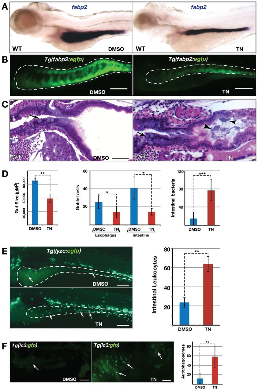
Intestinal pathologies of tunicamycin-treated wild-type larvae. (A) ISH with fabp2 shows smaller intestine in tunicamycin (TN)-treated larvae. (B) Confocal projections of Tg(fabp2:egfp) intestine (white outline) showing disrupted intestinal architecture of tunicamycin-treated larvae. (C) H&E-stained sections shows abnormal GCs (arrows) and increased luminal bacteria (arrowheads) in tunicamycin-treated larvae. (D) Bar charts showing reduced gut size, GC depletion and increased intestinal bacteria in tunicamycin-treated larvae (n≥10). (E) Confocal projection of Tg(lyzc:egfp) larval intestine shows increased macrophage aggregation (arrows) in tunicamycin-treated larvae (n=15). (F) Confocal projection of Tg(lc3:gfp) intestine shows increased autophagosomes (arrows) in tunicamycin-treated larvae (n=7). *P<0.05, **P<0.01, ***P<0.001. Scale bars: 100 µm (B,E); 20 µm (C); 5 µm (F).