
Image
|
Figure Caption
Fig. 2
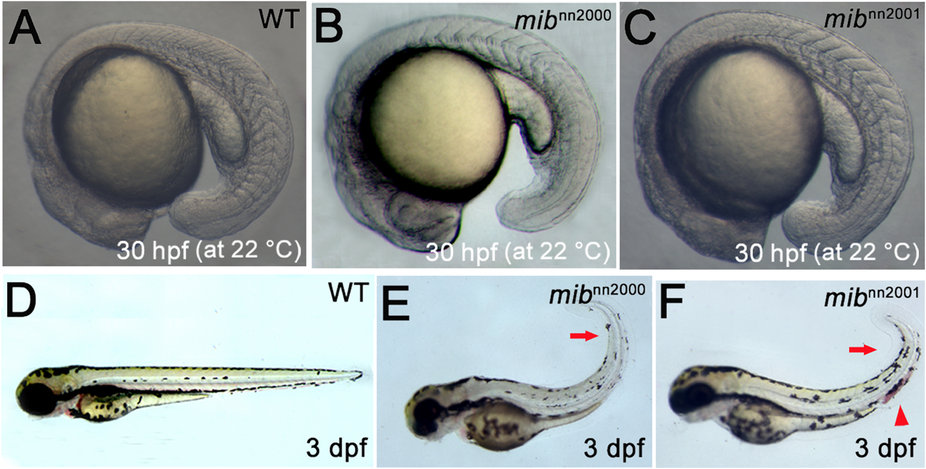
Morphological phenotypes of mibnn2000 and mibnn2001 mutants. A-C are the lateral views of 30 hpf embryos raised at 22 °C. While (A) WT embryos showed about 20 somites at 30 hpf, (B) mibnn2000 homozygotes showed about 11 recognizable somites and (C) mibnn2001 homozygotes showed about 14 somites. D-F are lateral views of 3 dpf embryos at 28.5 °C. Compared to (D) WT embryos, (E) mibnn2000 and (F) mibnn2001 mutants showed a curly tail (arrows), hemorrhage (arrowhead) and a reduction of tail pigmentation (more prominent in E).
Figure Data
Acknowledgments
This image is the copyrighted work of the attributed author or publisher, and
ZFIN has permission only to display this image to its users.
Additional permissions should be obtained from the applicable author or publisher of the image.
Full text @ Sci. Rep.