
IMAGE
Fig. 2
- ID
- ZDB-IMAGE-160513-4
- Publication
- Andrews et al., 2016 - Visualising apoptosis in live zebrafish using fluorescence lifetime imaging with optical projection tomography to map FRET biosensor activity in space and time
- All Figures
- Figures for Andrews et al., 2016
Image
|
Figure Caption
Fig. 2
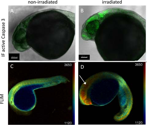
Validation of Caspase 3 biosensor activation following gamma irradiation using confocal microscopy. 24 hpf zebrafish embryos were irradiated (B, D) with 18 Gy from a 137Cs source or left untreated (A, C) and imaged at 3 hpi using confocal microscopy. (A, B) confocal fluorescence intensity images of TraNac embryos, either non-irradiated (A) or irradiated (B), fixed and labelled with an active Caspase 3 immunofluorescent antibody. (C, D) confocal fluorescence lifetime images of non-irradiated (C) and irradiated (D) Tg(Ubi : Caspase3bios) embryos at 3 hpi.
Acknowledgments
This image is the copyrighted work of the attributed author or publisher, and
ZFIN has permission only to display this image to its users.
Additional permissions should be obtained from the applicable author or publisher of the image.
Full text @ J. Biophotonics