
Fig. 6
- ID
- ZDB-IMAGE-160510-9
- Publication
- Lepanto et al., 2016 - Characterization of primary cilia during the differentiation of retinal ganglion cells in the zebrafish
- All Figures
- Figures for Lepanto et al., 2016
|
Fig. 6
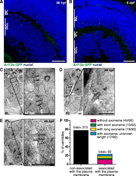
Primary cilia in maturing RGCs. a-b Arl13b-GFP transgenic embryos were fixed and imaged through confocal microscopy as whole-mounts (a, 48 hpf) or as cryosections (b, 5 dpf). Images shown are maximum intensity Z-projections of 1.5 µm (a) and 15 µm (b) confocal microscopy stacks. c-e TEM micrographs from retinas of 48 hpf embryos. In most of the cases we observed membrane-docked basal bodies without visible axonemes (c). Some cells showed basal bodies associated to long axonemes (d) or short axonemes with dilated tips (e). The white arrowhead indicates the basal body. f Comparison between the number of plasma membrane-associated and non-associated centrioles/centrosomes in the RGC layer of 48 hpf embryos (a total of 6 embryos were used in the quantification). The former were further classified in subtypes illustrated in the micrographs C, D and E. GCL: ganglion cell layer; IPL: inner plexiform layer; INL: inner nuclear layer; BL: basal lamina. Scale bars: A-B, 20 µm; C-E, 1 µm