
Fig. 2
- ID
- ZDB-IMAGE-160510-6
- Publication
- Lepanto et al., 2016 - Characterization of primary cilia during the differentiation of retinal ganglion cells in the zebrafish
- All Figures
- Figures for Lepanto et al., 2016
|
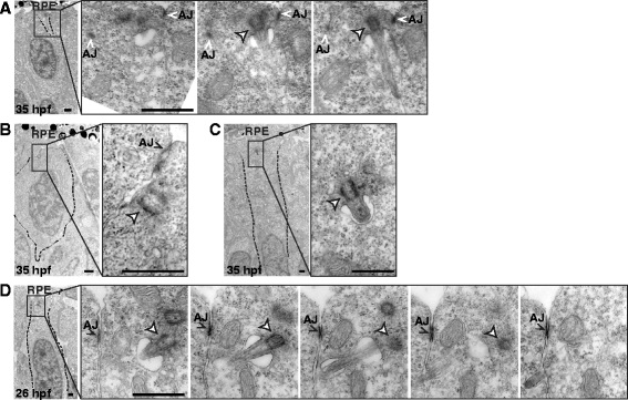
Fig. 2
Primary cilia emerging from the basolateral membrane of retinal neuroepithelial cells. TEM micrographs of retinas from 26 and 35 hpf embryos. a Low magnification (left) and high magnification serial sections (right) of a retina from a 35 hpf embryo. A primary cilium emerges from the basolateral membrane, as evidenced by the presence of adherent junctions (AJ). b Basal body without an axoneme, but closely associated with the basolateral membrane. c Basal body with a short axoneme inside a cytoplasmic vesicle. d Low magnification (left) and high magnification serial sections (right) of a retina from a 26 hpf embryo showing a primary cilium emerging from the basolateral membrane. White arrowhead: basal body; RPE: retinal pigment epithelium. Scale bars: A-D, 1 µm