
Fig. 7
- ID
- ZDB-IMAGE-160510-10
- Publication
- Lepanto et al., 2016 - Characterization of primary cilia during the differentiation of retinal ganglion cells in the zebrafish
- All Figures
- Figures for Lepanto et al., 2016
|
Fig. 7
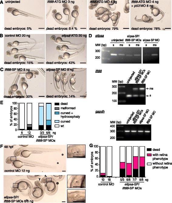
Evaluation of different morpholino oligomers for elipsa and ift88 knock-down. a-c External phenotype of 48 hpf embryos injected with different morpholinos targeting the ciliary proteins ift88 and elipsa; translational-blocking morpholinos: ift88-ATG MO (a), elipsa-ATG MO (b); splice-blocking morpholinos: ift88-SP or elipsa-SP (c). d RT-PCR analysis of elipsa and ift88 mRNA levels in 35 hpf embryos either injected with ift88-SP (6 ng) or elipsa-SP (6 ng) alone or as a combination (6 ng each) (“s”: spliced mRNA form; “ms”: mis-spliced mRNA form). Gapdh mRNA was used as a control. e Evaluation of the external phenotype of 48 hpf embryos injected with different amounts of the combination of splice-blocking morpholinos (elipsa-SP/ift88-SP MOs), where “malformed” refers to morphological alterations not compatible with a classic “ciliary phenotype”. f Main characteristics of the external phenotype of double morphants at the dose used in the rest of the study. The black rectangle marks the position of the otic vesicle in the low magnification images, magnified in the insets. The arrowheads indicate embryos with enlarged brain ventricles. g Quantification of the percentage of atoh7:gap-GFP embryos injected with different amounts of control MO or elipsa-SP/ift88-SP MOs displaying reduced size of the RGC layer (“retina phenotype”), at 48 hpf. Scale bars: A-C, 500 µm; F, 200 µm