
|
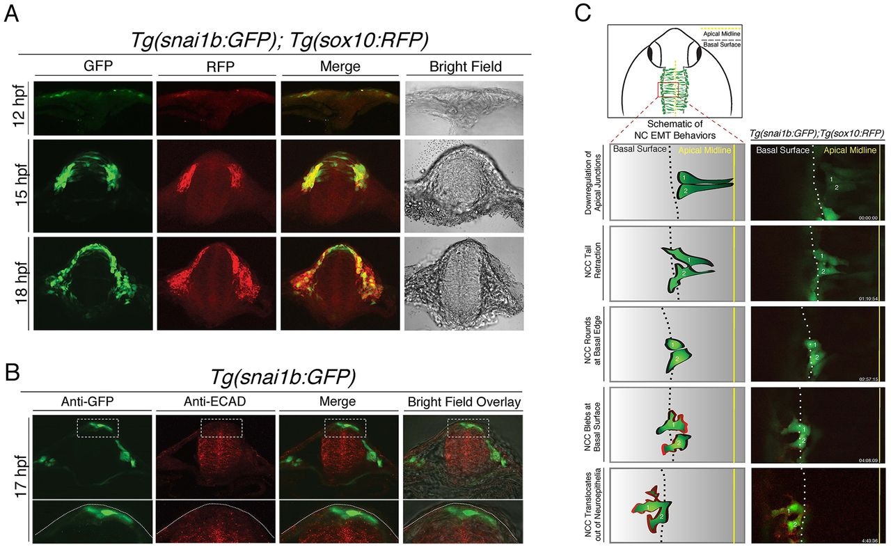
Fig. 2
Tg(snai1b:GFP) labels dorsal neural tube progenitors that display morphological cell behaviors of EMT. (A) Transverse hindbrain sections in double-transgenic Tg(snai1b:GFP); Tg(sox10:RFP) embryos at 12, 15 and 18hpf (single confocal z plane, 40×) showing that snai1b-driven GFP is expressed in dorsal neural tube progenitor cells at 15hpf, whereas both GFP and sox10-driven RFP are expressed in cells adjacent to the neural tube and migrating NC cells. (B) Transverse hindbrain sections of Tg(snai1b:GFP) embryos at 17hpf processed for immunofluorescence analysis of Cdh1 (ECAD) and GFP. Bottom row displays higher-magnification views of boxed regions, and show decreased ECAD levels in GFP-positive cells compared to ventral neural tube. Dotted lines in the bottom panel outline the dorsal neural tube. (C) Schematic illustrating the imaging area (top) and cell delamination behavior (bottom left) derived from confocal time-lapse images of Tg(snai1b:GFP); Tg(sox10:RFP) embryos (bottom right), which captured two cells delaminating out of the neuroepithelium to produce sox10-positive NC cells (~14hpf to 18hpf).