
Image
|
Figure Caption
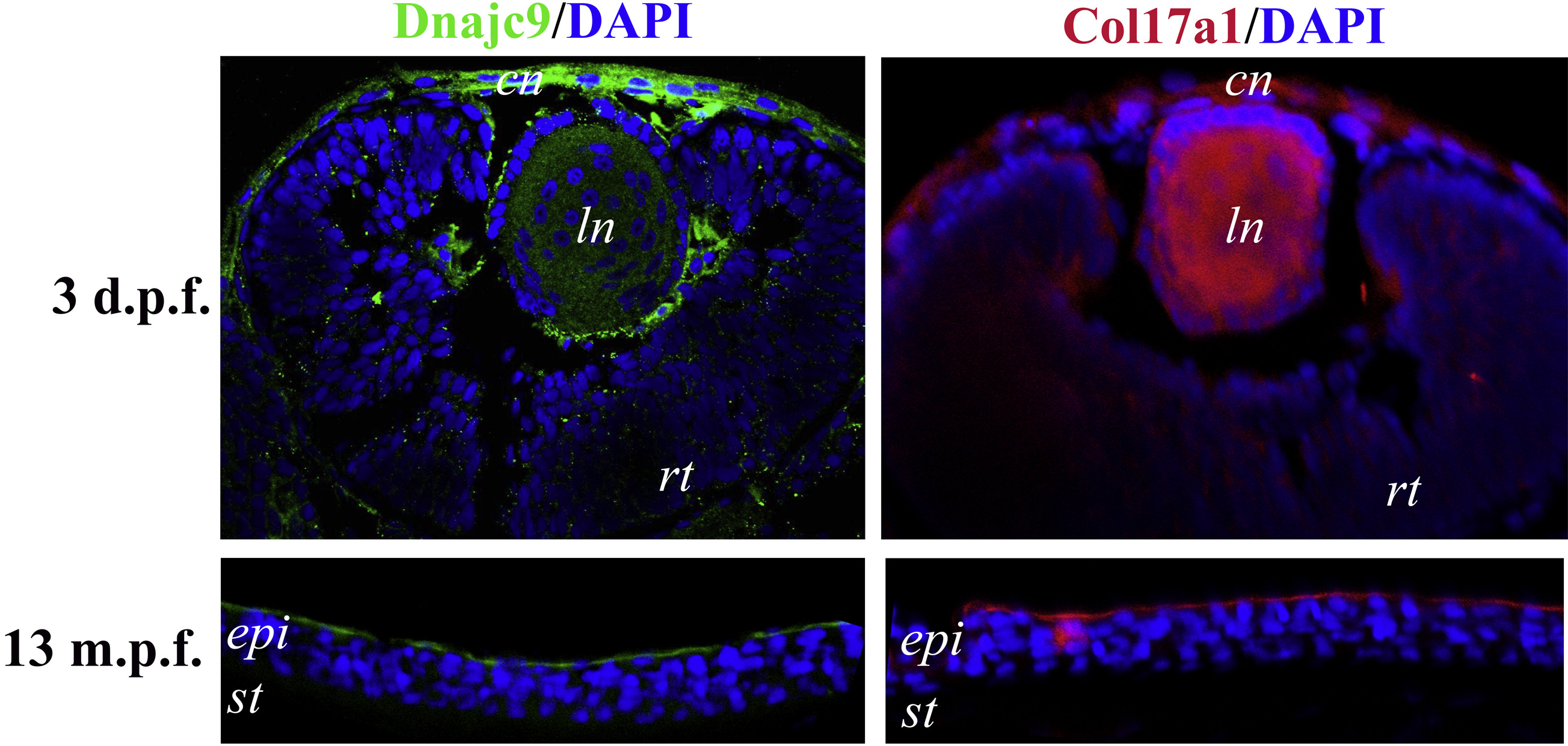
Fig. 5
Immunohistochemistry staining results for Dnajc9 and Col17a1 protein in zebrafish cornea. Both Dnajc9 (green) and Col17a1 (red) proteins are present in the dual cell layers of the developing zebrafish cornea (3 days after fertilization [d.p.f.]). In the adult zebrafish cornea (13 months after fertilization [m.p.f.]), Dnajc9 and Col17a1 expression is restricted to the external surface membrane of the superficial epithelial cells. 4′,6-Diamidino-2-phenylindole staining (blue) indicates cell nuclei. cn = Cornea; epi = epithelium; ln = lens; rt = retina; st = stroma.
Figure Data
Acknowledgments
This image is the copyrighted work of the attributed author or publisher, and
ZFIN has permission only to display this image to its users.
Additional permissions should be obtained from the applicable author or publisher of the image.
Full text @ Ophthalmology