
Fig. S2
- ID
- ZDB-IMAGE-160407-11
- Publication
- Zhang et al., 2016 - A Naturally-Derived Compound Schisandrin B Enhanced Light Sensation in the pde6c Zebrafish Model of Retinal Degeneration
- All Figures
- Figures for Zhang et al., 2016
|
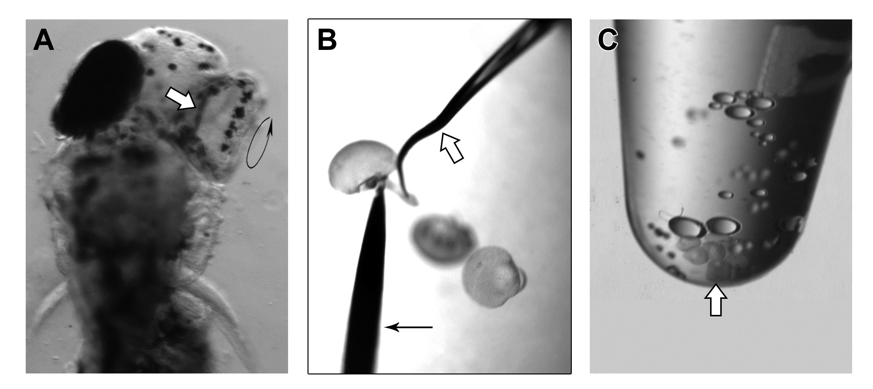
Fig. S2
Microdissection of larval retina for whole-mount immunostaining.In this study, larval retinas were microdissected from 6-dpf larvae. First, the scleral region just outside the circumference of the pupil was severed from the lateral side of the larvae (black circular arrow in A) by a fine hook created and bent from a chemically-etched tungsten needle [39]. An example of this needle is indicated by the white arrow in (B). In the same figure, the black arrow indicates an insect pin of size 000 (Fine Science Tools, Foster City, CA). Three 6-dpf retinas are shown in the figure to give a reference of the relative size. After severing the scleral attachment from the lateral side (circular arrow in A), the RPE-attached retinas could be easily detached from the sclera by a gentle push from the medial side (A, white arrow). These RPE-retinas were then treated with acetone, which would further detach the RPE from the retinas. The detached RPE was removed from the retinas by the chemically-etched tungsten needle. (C) Finally, the dissected retinas (indicated by the white arrow) were collected in a microcentrifuge tube for downstream immunostaining procedure.