
Fig. 4
- ID
- ZDB-IMAGE-160331-3
- Publication
- McMillen et al., 2016 - A Sawtooth Pattern of Cadherin 2 Stability Mechanically Regulates Somite Morphogenesis
- All Figures
- Figures for McMillen et al., 2016
|
Fig. 4
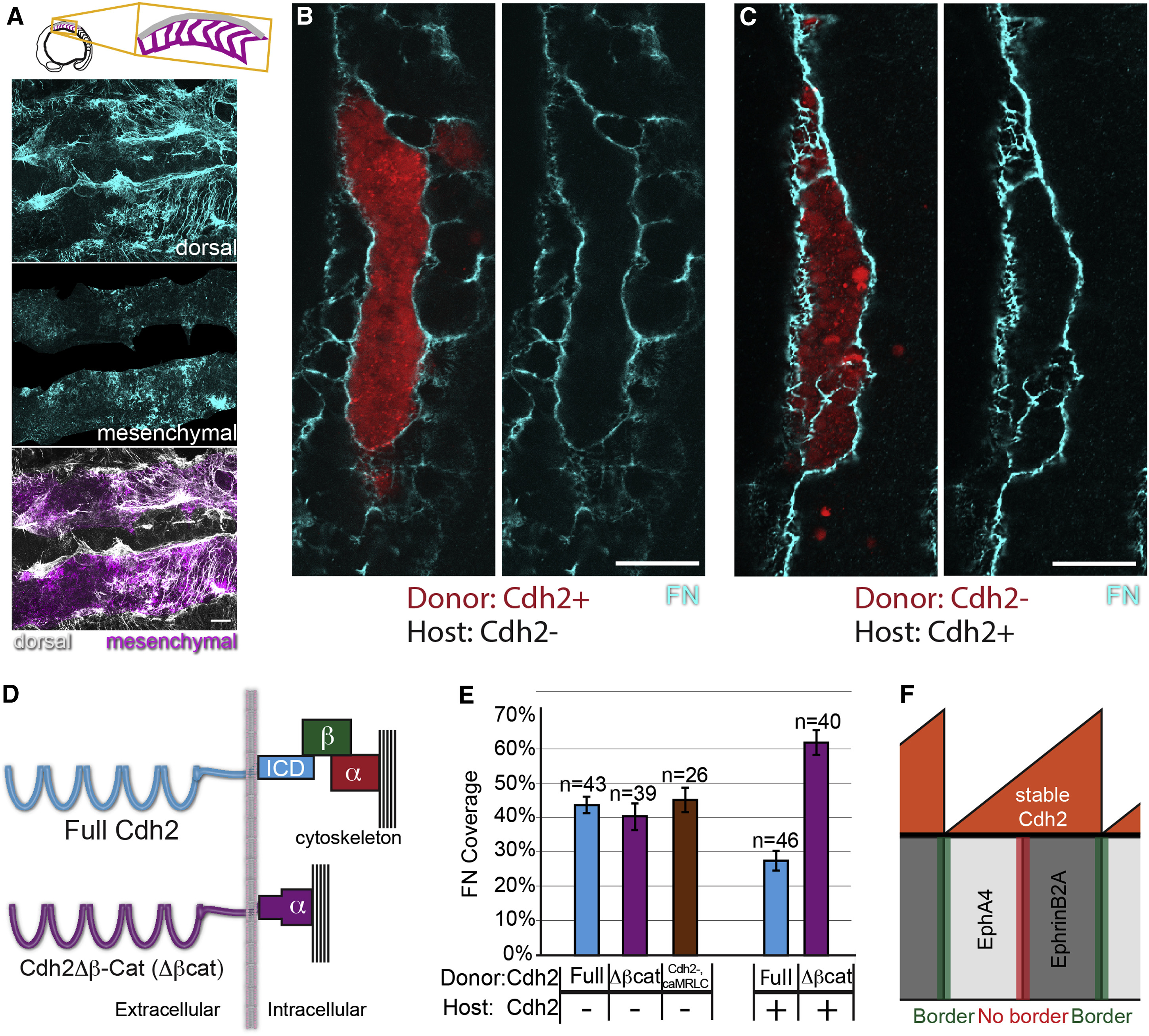
Cdh2 Bimodally Regulates ECM Assembly through Distinct Molecular Mechanisms
(A) MZitgα5-/-;cdh2mo embryos exhibit decreased dorsal ECM assembly and increased ectopic mesenchymal ECM.
(B and C) In mosaic embryos, mesenchymal ECM forms in either Cdh2– hosts (B) or donor clones (C), but not among Cdh2+ neighboring cells.
(D) Schematic of the Cdh2Δβ-Cat (Δβcat) construct in which Cdh2 is fused directly to α-catenin, thus mediating adhesion without binding β-catenin.
(E) Δβcat stimulates FN matrix assembly to the same extent as full-length Cdh2 (p > 0.25) but does not inhibit FN matrix assembly (p < 0.0001). caMRLC also stimulates FN matrix assembly (p < 0.0001) to the same extent as full-length Cdh2 and Cdh2Δβ-Cat (p > 0. 25). n, number of clones examined. Mean values indicated ± SEM. Scale bars, 20 µm. p values were determined by t test.
(F) A model schematic showing graded stable Cdh2 levels inhibiting boundary formation at Eph/ephrin interfaces within somites while differential levels of stable Cdh2 promote boundary formation at Eph/ephrin interfaces between somites.
See also Figure S3.