
|
Fig. 1
Lipid Distribution and Metabolic Gene Expression throughout Embryogenesis
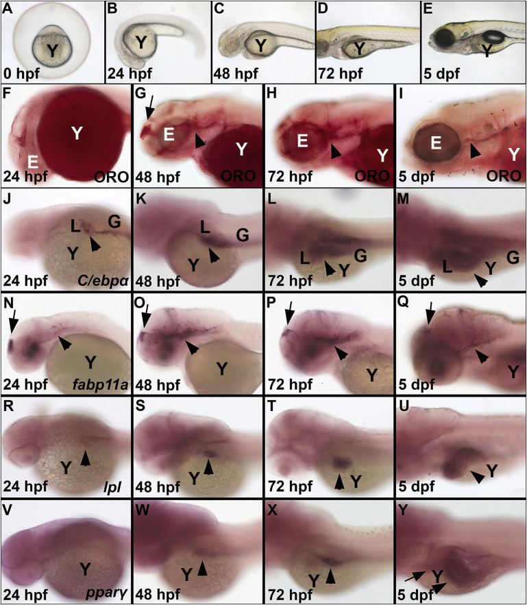
(A-E) Bright field images show yolk utilization from 0 hpf to 5 dpf.
(F-I) ORO staining shows that there were no deposits of neutral lipid in the head of the embryo at 24 hpf (F), but lipid was present at 48 hpf in the forebrain (arrow, G) and around the eye and underneath the otic vesicle (arrowhead, G), at 72 hpf around the eye (arrowhead, H), and at 5 dpf at a low amount (I).
(J) c/ebpα expression was present at 24 hpf in the developing liver and gut (arrowhead).
(K-M) c/ebpα expression increased in these organs from 48 hpf to 5 dpf (arrowhead).
(N-Q) fabp11a expression was located in the forebrain (arrow), in the eye and in the hindbrain (arrowhead) from 24 hpf to 5 dpf.
(R-U) lpl was present in the developing liver (arrowhead) from 24 to 5 dpf.
(V) pparγ was not detected in the embryo at 24 hpf.
(W and X) At 48 and 72 hpf, pparγ was expressed in the developing gut (arrowheads).
(Y) At 5 dpf, expression increased in the liver (arrow) and gut (arrowhead) and was present in the swim bladder. E, eye; G, gut; L, liver; Y, yolk.
See also Figure S1.