
Image
|
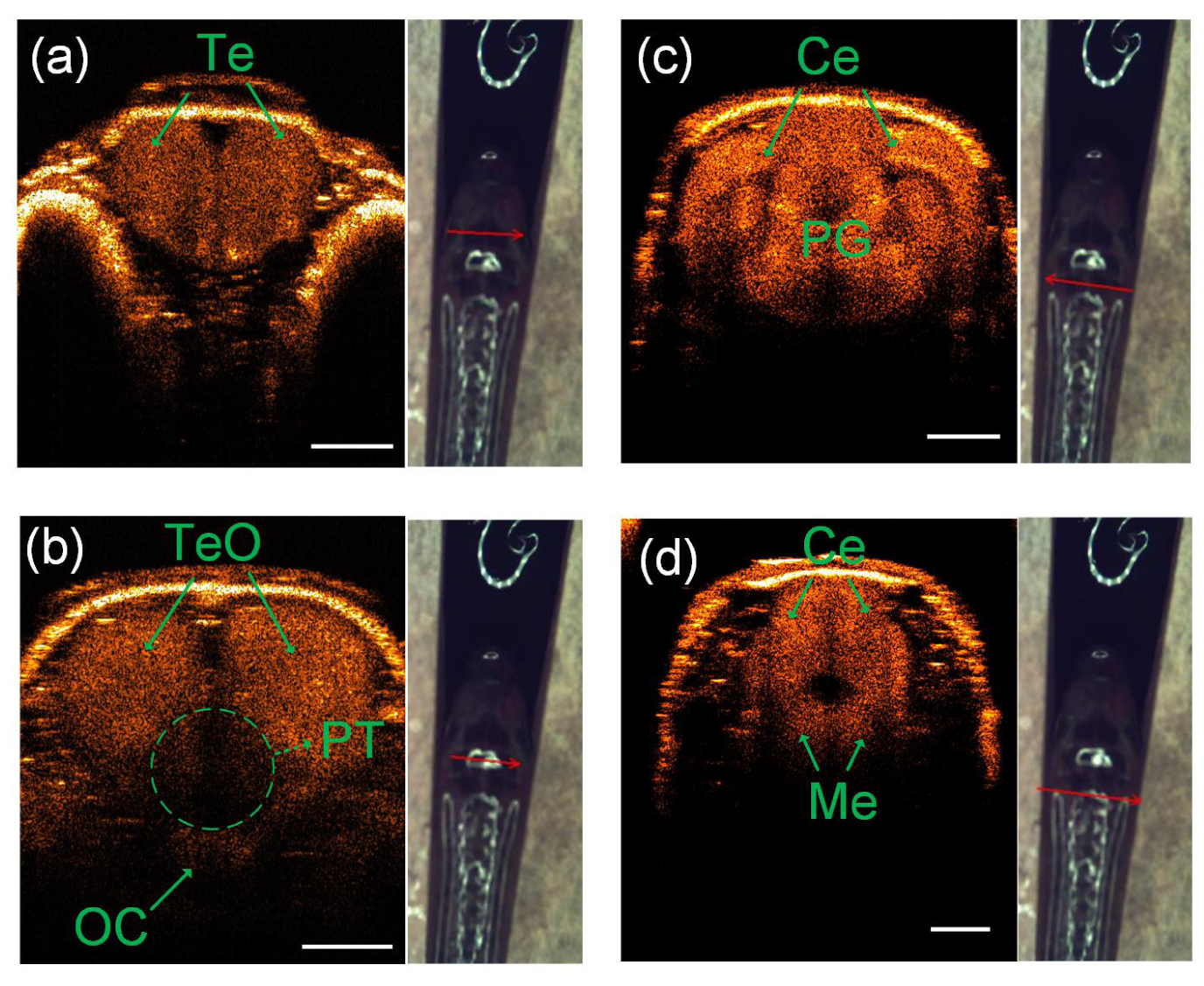
Figure Caption
Fig. 3
Four different coronal SD-OCT images [(a) to (d)] at four different labeled positions on the head of the same adult zebrafish (see arrow locations and direction): OC, optic commissure; Te, telencephalon; TeO, tectum opticum; Ce, cerebellum; Me, medulla; PG, preglomerular complex; PT, posterior tuberculum. The scale bar is 500 µm.
Acknowledgments
This image is the copyrighted work of the attributed author or publisher, and
ZFIN has permission only to display this image to its users.
Additional permissions should be obtained from the applicable author or publisher of the image.
Full text @ Biomed. Opt. Express