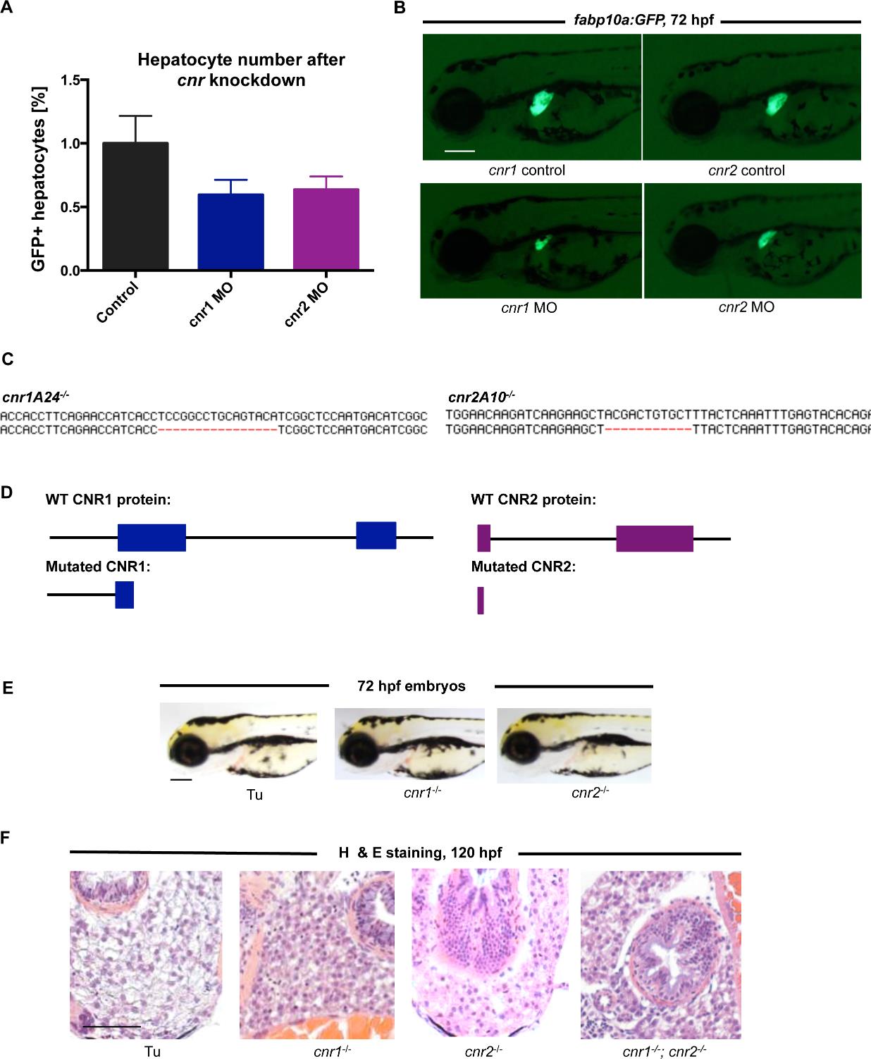
Fig. S2
Characterization of cannabinoid receptor mutants and morphants
A) FACS quantification of fabp10a:GFP embryos injected with cnr1 and cnr2 morpholinos. Morphant embryos at 72hpf show a decreased number of GFP-positive hepatocytes.
Mean±s.e.m., n=5 groups of 10 pooled embryos; p=0.18, one-way ANOVA analysis.B) Fluorescent micrographs showing that morpholino knockdown of cnr1 or cnr2 in fabp10a:GFP reporter fish leads to smaller livers at 72hpf. Scale bar = 0.2 mm.
C) Depiction of wild type and mutant sequence of the TALEN target region in the cnr1 and cnr2 genes.
D) Small deletions in the DNA sequence lead to an early stop codon in the first exon of both cnr1 and cnr2.
E) Brightfield images of cnr1-/- and cnr2-/- zebrafish at 72hpf reveal no gross anatomical abnormalities or significant developmental delay compared to wild type animals. Scale bar = 0.2 mm.
F) H & E staining of transverse sections in 120hpf larvae reveals similarly abnormal liver histology in cnr1-/-; cnr2-/- double mutants compared to histology found in the cnr1-/- and cnr2-/- single mutants shown in Figure 3A. Scale bar = 0.1 mm.

