
|
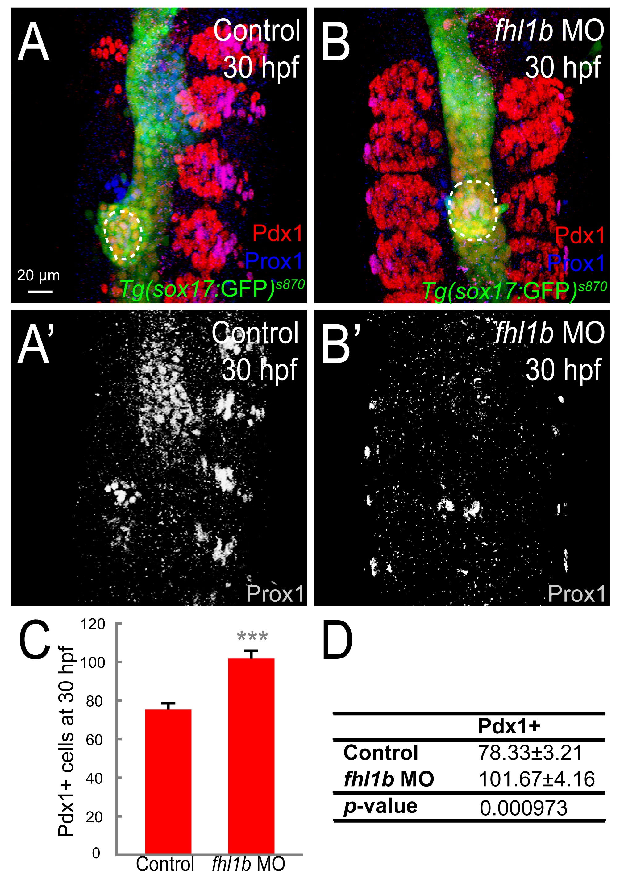
Fig. S4
Loss of Fhl1b activity compromises liver specification and enhances induction of Pdx1-positive cells in the dorsal pancreatic bud.
(A-B′) Confocal images of Tg(sox17:GFP)s870 control embryos (A and A2) and fhl1b morphants (B and B2) at 30 hpf, stained for Pdx1 (red; dorsal pancreatic bud is outlined by white dotted circles) and Prox1 (blue in A and B; grey in A′ and B′). The somites are also Pdx1 positive. Compared to control embryos (A and A2), in fhl1b morphants (B and B2), the Pdx1 expression domain in the dorsal pancreatic bud was expanded, while the Prox1 expression domain was significantly reduced. (C-D) Quantification of the number (mean±SD) of Pdx1-positive cells in the pancreas at 30 hpf. Cells in 20 planes of confocal images from 5 individual embryos were counted. Asterisks indicate statistical significance: ***, P < 0.001. A-B′, confocal projection images, ventral views, anterior to the top. Scale bar, 20 µm.