
Fig. S2
- ID
- ZDB-IMAGE-160316-25
- Publication
- Yoshimatsu et al., 2016 - Presynaptic partner selection during retinal circuit reassembly varies with timing of neuronal regeneration in vivo
- All Figures
- Figures for Yoshimatsu et al., 2016
|
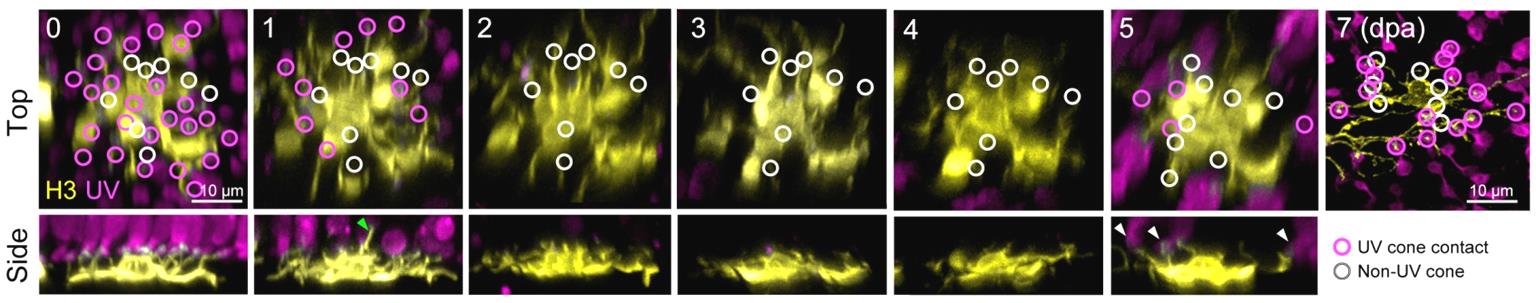
Fig. S2
H3 HCs retract dendritic tips as UV cones die, and rewire with the regenerated population of UV cones.
Two-photon time-lapse imaging of a H3 HC in the background of Tg(sws1:nfsB-mCherry) beginning at 5 dfp and subsequently after cone ablation and regeneration. Circles indicate the locations of dendritic tips contacting UV cones (magenta) or dendritic tips that were not associated with UV cones (white, non-UV cones). Green arrowhead (1 dpa) points to a dendrite sprouting into the photoreceptor layer. White arrowheads (5 dpa) indicating new dendritic contacts with newly generated UV cones. dpa, days postablation.