
Fig. 1
- ID
- ZDB-IMAGE-160316-1
- Publication
- He et al., 2016 - Histone deacetylase 1 is required for the development of the zebrafish inner ear
- All Figures
- Figures for He et al., 2016
|
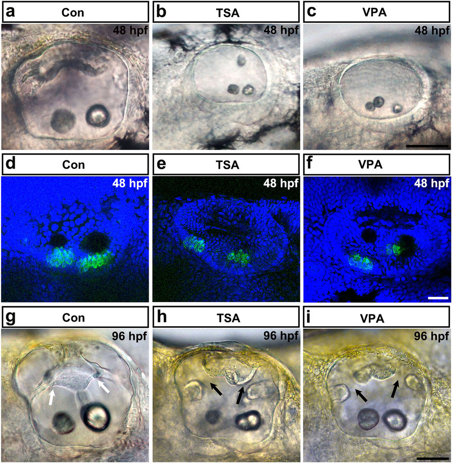
Fig. 1
HDAC inhibitor treatment induces otic abnormalities and reduces the number of hair cells in the inner ear.
(a-c) The morphology of the otoliths in control embryos and in embryos treated with TSA or VPA from 12 hpf onwards. HDAC inhibitor treatment caused morphant otolith defects at 48 hpf of development. (d-f) Images of hair cells expressing GFP in control, TSA-treated, and VPA-treated transgenic zebrafish at 48 hpf. Hair cells were detected by brn3c:gfp expression. HDAC inhibitor treatment reduced hair cell numbers in the inner ear. (g-i) The morphology of the semicircular canals in control (g), TSA-treated (h), and VPA-treated (i) zebrafish. HDAC inhibitor treatment induced aberrant semicircular canal formation. White arrows mark fused pillars, and black arrows mark unfused projections. All images are lateral views with the anterior to the left and the dorsal side up. Scale bars are 50 µm (a-c,g-i) and 20 µm (d-f).