
|
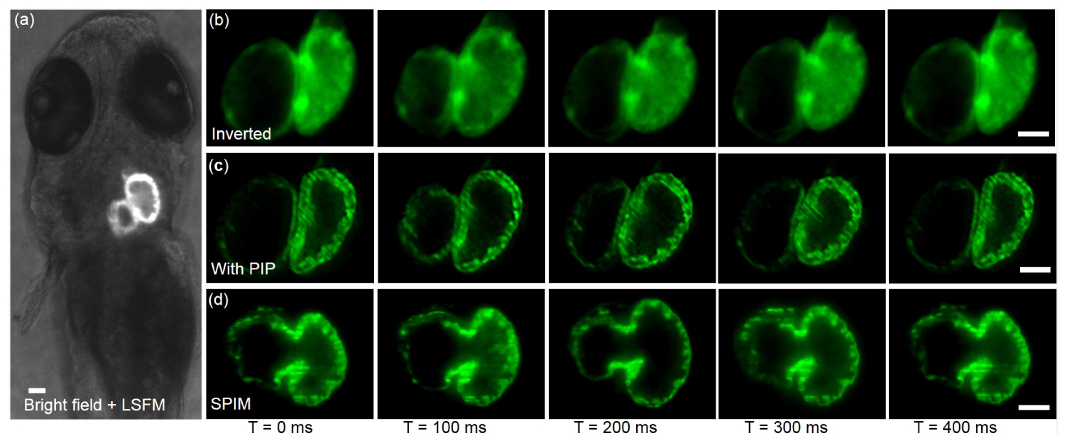
Fig. 4
Contrast-enhanced light sheet imaging of beating embryonic zebrafish heart by PIP mounted microscope. (a) Light sheet sectioning inside a live embryonic zebrafish heart (4 d.p.f., Tg: (cmlc: gfp)). PIP enables the selective plane illumination on specific region of interest of the beating heart. (b) and (c) show the image comparison between regular inverted microscope and PIP mounted inverted microscope. The dynamic inner and outer boundaries of the beating heart can be clearly resolved when PIP is added. (d) shows the control heart images (same stage embryo) from a home-built, standard SPIM system with identical illumination and detection settings of PIP imaging. Scale bars in all images are 50 µm.