
|
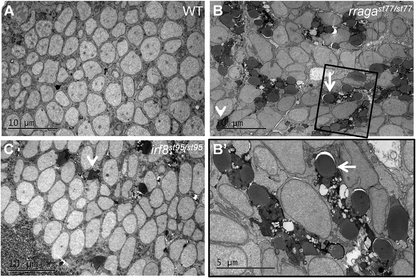
Fig. 4
Abnormal Vacuolar Organelles in Microglia and Uncleared Apoptotic Neurons in rragast77/st77 Mutants
(A–C) Transmission electron micrographs of the dorsal midbrain of (A) WT, (B and B′) rragast77/st77, and (C) irf8st95/st95 larvae at 4 dpf. Boxed region in (B) is shown at higher magnification in (B′). Microglia in the rragast77/st77 mutants have a striking accumulation of vacuolar organelles that appear to contain undigested material (white arrows). Microglia were absent in the irf8 mutant. In both mutants, uncleared corpses of apoptotic neurons are present (white arrowheads). These phenotypes also were evident in mutants stained with acridine orange ( Figure S3). Toluidene blue-stained sections of the same larvae are shown in Figure S4. Larvae were genotyped by PCR from tail biopsies collected immediately prior to fixation.