
Fig. 3
- ID
- ZDB-IMAGE-160308-19
- Genes
- Publication
- Nguyen et al., 2016 - Development of a conditional liver tumor model by mifepristone-inducible Cre recombination to control oncogenic kras(V12) expression in transgenic zebrafish
- All Figures
- Figures for Nguyen et al., 2016
|
Fig. 3
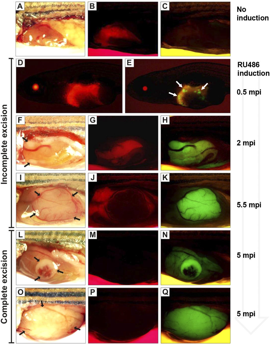
Mosaic pattern of Cre-mediated activation of EGFP-krasV12 in transgenic fish.
Brightfield and corresponding fluorescence images of representative Triple-Tg fish are shown in the same rows. (A–C) Without induction, Triple-Tg fish showing normal liver morphology (A) with mCherry (B) but not EGFP-krasV12 (C). (D–K) Induced Triple-Tg fish at 1-month-old expressing both mCherry (D,G,J) and EGFP-krasV12 (E,H,K) in the liver, indicating the occurrence of incomplete Cre excision. After induction, many subsets of EGFP-positive liver cells were observed in 1.5-month-old fish (D,E). Liver tumors expressing EGFP developed in a 2.5-month-old (F,G,H) and 6-month-old Triple-Tg fish (I,J,K). (L–Q) Complete excision of the LChL cassettes observed in induced Triple-Tg fish at 6-month-old with the formation of large liver tumors (L,O) only expressing EGFP-krasV12 (N,Q) and no detectable mCherry fluorescence (M,P). Liver tumors are denoted by arrows.