Fig. S7
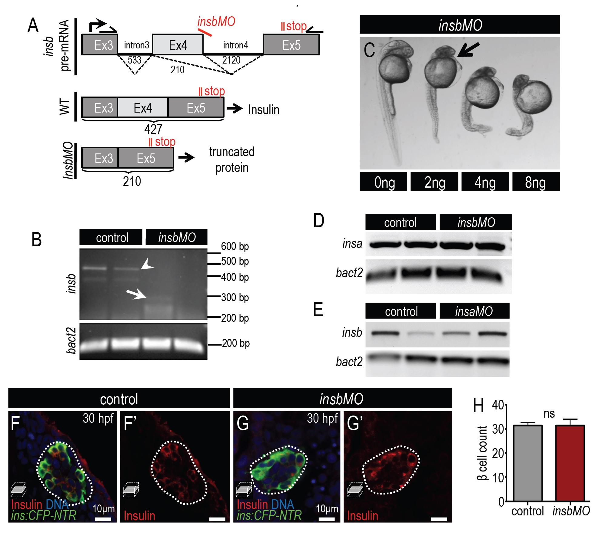
Knockdown of insb results in severe developmental defects. (A) Diagram illustrating insb pre-mRNA structure in wild type and insbMO-injected cells. insbMO targets the exon 4-intron 4 boundary of preproinsulin-b mRNA and promotes the deletion of exon 4. Primers complementary to exon 3 and exon 5 amplify the differentially spliced cDNA products. (B) Agarose gel showing amplified insb PCR product from 2 dpf control and insbMO-injected embryos. Note that a PCR product with a size of 427 bp was detected in control embryos (arrowhead) but not insbMO injected embryos. A weak product of 210 bp can be detected in insbMO-injected embryos (arrow). β actin was used as an internal control. (C) Dose dependent phenotypes at 48 hpf after insbMO injection. Arrow indicates head defect first observed at 2 ng insbMO dose. (D,E) RT-PCR analysis of cDNA in 30 hpf insbMO-injected (D) and insaMOinjected (E) embryos. Neither insbMO nor insaMO affected the splicing or expression of the mRNA of the respective paralog. (F-G′) Merged and single channel confocal planes of 24 hpf control (F,F′) and insbMO-injected (G,G′) islets of Tg(ins:CFP-NTR) embryos stained for CFP (β cells, green) and insulin (red). (H) Quantification of ins:CFP-NTR+ β cells in 24 hpf control (n=5) and insbMO-injected embryos (n=6). Student′s t-test was used to determine significance in H.
Reprinted from Developmental Biology, 409(2), Ye, L., Robertson, M.A., Mastracci, T.L., Anderson, R.M., An insulin signaling feedback loop regulates pancreas progenitor cell differentiation during islet development and regeneration, 354-69, Copyright (2016) with permission from Elsevier. Full text @ Dev. Biol.

