
|
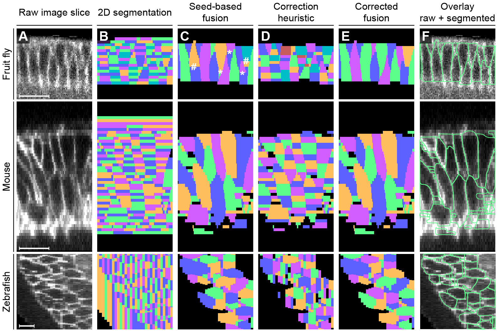
Fig. S3
Related to Figure 1. Efficient seed-based fusion of 2D segments to 3D cell shapes.
Illustration of final segment fusion steps for 3D cell shape segmentation in cell membrane image data (A, xz-image is shown, i.e. an image slice oriented parallel to the microscope’s optical detection axis). The initial slice-based segmentation of the 2D morphological watershed operator (B) and the extracted seed segments are combined to reconstruct 3D cell shapes (C). 2D segments touching a seed segment are labeled with the unique seed identifier. Based on segment similarity across slices, 2D segments touching one of the labeled initialization segments are then iteratively merged to form complete 3D cell shapes, starting with the highest-scoring segments. In some cases, the 3D segmentation results (C) can be further improved by one of the two proposed fusion heuristics. Segments with few slices (indicated by * in panel (C)) are fused to their closest neighbor if the specimen-dependent maximum volume constraint is not violated by this merge. If the fusion of segments is suggested by the similarity-based fusion heuristic (D), two subcellular compartments produced in the initial seed-based segmentation (indicated by # in panel (C)) are fused to obtain the final 3D segmentation (E, F). Both fusion heuristics simply assign the same unique identifier to those segments that should be merged, thus producing a single corrected 3D cell shape. For specimens with diverse cell sizes, the fusion heuristics might not be able to further improve results (see e.g. the almost identical results obtained for the zebrafish and mouse panels shown before (C) and after (E) application of the fusion heuristics; see also Part 1 of Supplemental Experimental Procedures). We note that most of the small fragments visible in (E) and (F) are not over-segmentation artifacts but rather cell cross-sections belonging to cells whose centers are located outside the xz-slice shown here. Scale bar, 20 µm.
Reprinted from Developmental Cell, 36, Stegmaier, J., Amat, F., Lemon, W.C., McDole, K., Wan, Y., Teodoro, G., Mikut, R., Keller, P.J., Real-Time Three-Dimensional Cell Segmentation in Large-Scale Microscopy Data of Developing Embryos, 225-240, Copyright (2016) with permission from Elsevier. Full text @ Dev. Cell