
IMAGE
Fig. S3
- ID
- ZDB-IMAGE-160229-18
- Publication
- Cao et al., 2016 - Single epicardial cell transcriptome sequencing identifies Caveolin-1 as an essential factor in zebrafish heart regeneration
- All Figures
- Figures for Cao et al., 2016
Image
|
Figure Caption
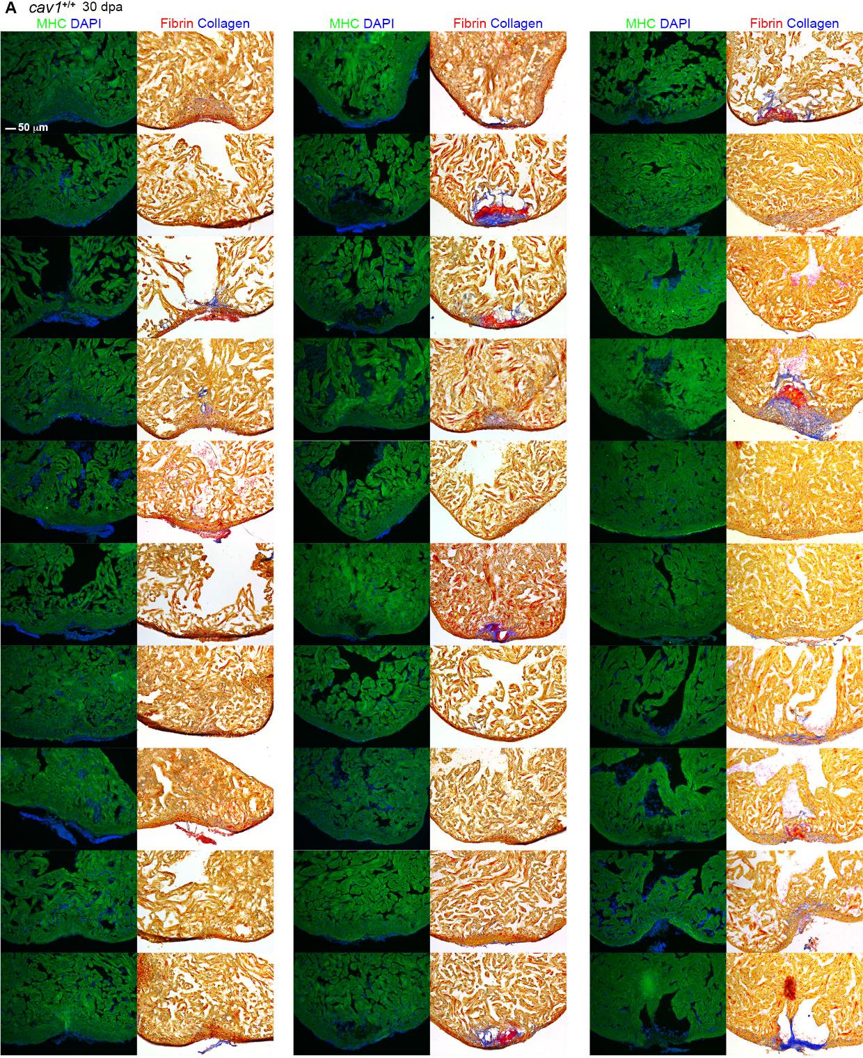
Fig. S3
Representative sections from each of 91 hearts of wild-types (A), cav1 heterozygotes (B), and cav1 homozygotes (C), at 30 dpa. Sections from uninjured cav1 homozygous mutant hearts are shown in (D). Ventricular sections are stained for MHC (green) to denote cardiac muscle. The same sections in left panels are stained with acid fuchsin orange G to characterize nonmuscle components in the injuries (right; blue for collagen, red for fibrin).
Acknowledgments
This image is the copyrighted work of the attributed author or publisher, and
ZFIN has permission only to display this image to its users.
Additional permissions should be obtained from the applicable author or publisher of the image.
Full text @ Development