
IMAGE
Fig. 8
- ID
- ZDB-IMAGE-160229-14
- Publication
- Cao et al., 2016 - Single epicardial cell transcriptome sequencing identifies Caveolin-1 as an essential factor in zebrafish heart regeneration
- All Figures
- Figures for Cao et al., 2016
Image
|
Figure Caption
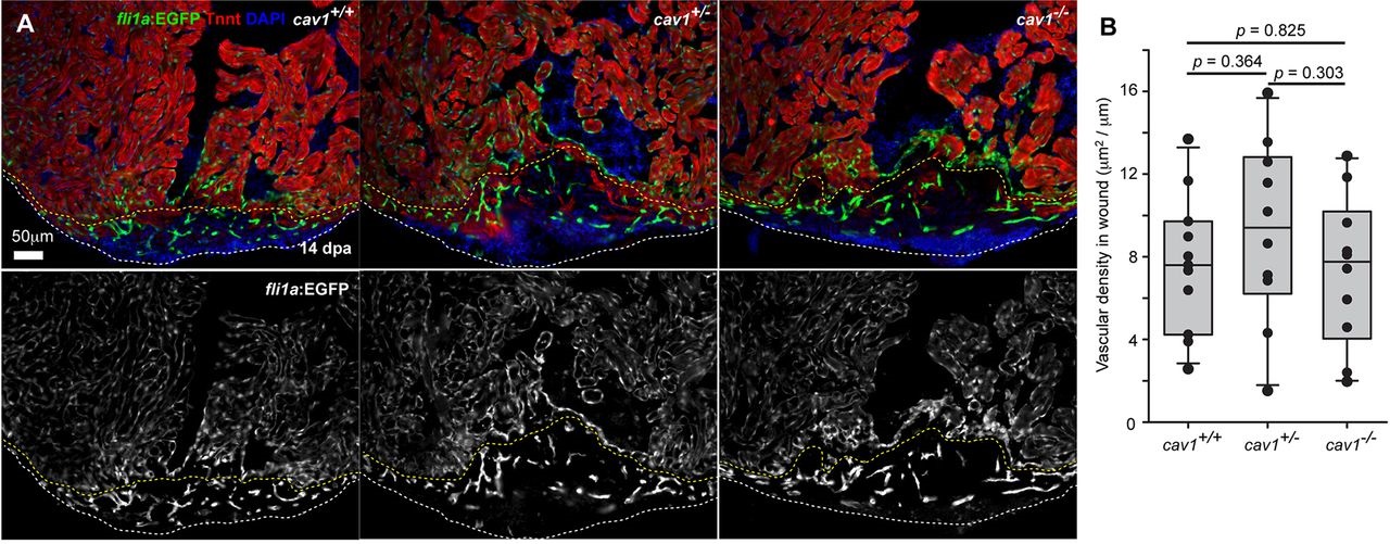
Fig. 8
Cardiac injury vascularization in cav1 mutants. (A) Sections of injured hearts of fli1a:EGFP fish in three cav1 genotypes at 14dpa, stained for TnnT (red). White dashed lines indicate wound edges, and yellow dashed lines delineate vascularized area from endocardial area. (B) Quantification of EGFP+ vascular endothelial area in wounds with respect to wound edge lengths. Student′s t-test; n=11 for wild type, n=10 for heterozygotes, and n=10 for homozygotes.
Acknowledgments
This image is the copyrighted work of the attributed author or publisher, and
ZFIN has permission only to display this image to its users.
Additional permissions should be obtained from the applicable author or publisher of the image.
Full text @ Development