
IMAGE
Fig. 7
- ID
- ZDB-IMAGE-160229-13
- Publication
- Cao et al., 2016 - Single epicardial cell transcriptome sequencing identifies Caveolin-1 as an essential factor in zebrafish heart regeneration
- All Figures
- Figures for Cao et al., 2016
Image
|
Figure Caption
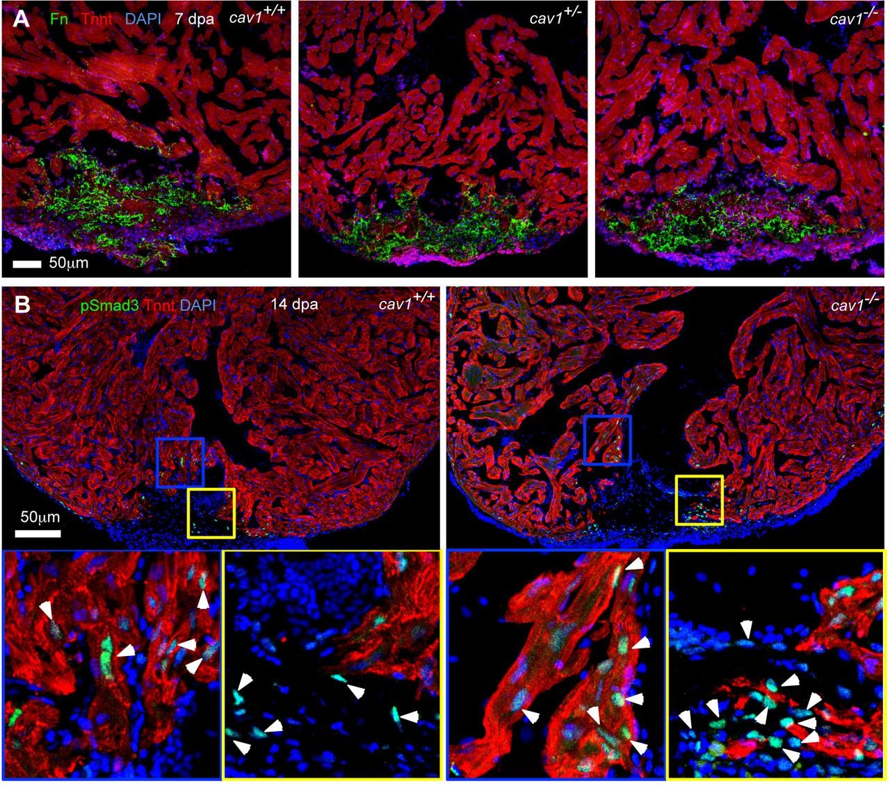
Fig. 7
Fibronectin deposition and Tgfβ signaling activation in cav1 mutants. (A) Section images of injured ventricles from the three cav1 genotypes at 7dpa. Sections are stained for Tnnt (red) and fibronectin (Fn, green). (B) Sections of injured ventricles at 14dpa are stained for Tnnt (red) and pSmad3 (green). The boxed regions are enlarged to show details. Arrowheads indicate pSmad3-positive CMs (blue box, Tnnt+) and non-CMs (yellow box, Tnnt -).
Acknowledgments
This image is the copyrighted work of the attributed author or publisher, and
ZFIN has permission only to display this image to its users.
Additional permissions should be obtained from the applicable author or publisher of the image.
Full text @ Development