
Fig. 6
- ID
- ZDB-IMAGE-160229-12
- Publication
- Cao et al., 2016 - Single epicardial cell transcriptome sequencing identifies Caveolin-1 as an essential factor in zebrafish heart regeneration
- All Figures
- Figures for Cao et al., 2016
|
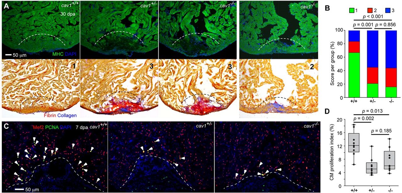
Fig. 6
Cav1 is required for zebrafish heart regeneration. (A) cav1 heterozygous and homozygous mutants displayed massive defects in muscle regeneration (top) and fibrin/collagen retention (bottom). Section images of ventricles at 30dpa are stained for Myosin heavy chain (MHC, green) to indicate cardiac muscle. Dashed line indicates plane of resection. The same sections as in the top panels are stained with Acid Fuchsin-Orange G to characterize non-muscle components in the injuries (bottom; blue for collagen, red for fibrin). Twenty-two of 29 ventricles from cav1 heterozygotes and 27 of 32 from cav1 homozygotes showed obvious areas of missing myocardium and prominent scar deposition, compared with 8 of 30 ventricles from wild-type siblings. P<0.001 (Fisher′s exact test) for both heterozygous and homozygous hearts. The numbers 1-3 indicates scores as in B. (B) Quantification of regeneration in cav1 mutants and wild-type siblings at 30dpa. Representative hearts, such as those shown in A, were scored for regeneration: 1, complete regeneration of a new myocardial wall; 2, partial regeneration; 3, a strong block in regeneration. Data indicate the percentage of total hearts represented by each score for each genotype, and were analyzed by Chi-square tests. (C) Section images of injured ventricles from the three cav1 genotypes at 7dpa. Sections are stained for Mef2 (red) and PCNA (green). Arrowheads indicate Mef2+ PCNA+ nuclei. Dashed line indicates plane of resection. (D) Quantification of CM proliferation indices in 7dpa ventricles. For each group, nine fish were assessed, and data were analyzed using Mann–Whitney tests.