
Fig. 4
- ID
- ZDB-IMAGE-160229-10
- Genes
- Publication
- Cao et al., 2016 - Single epicardial cell transcriptome sequencing identifies Caveolin-1 as an essential factor in zebrafish heart regeneration
- All Figures
- Figures for Cao et al., 2016
|
Fig. 4
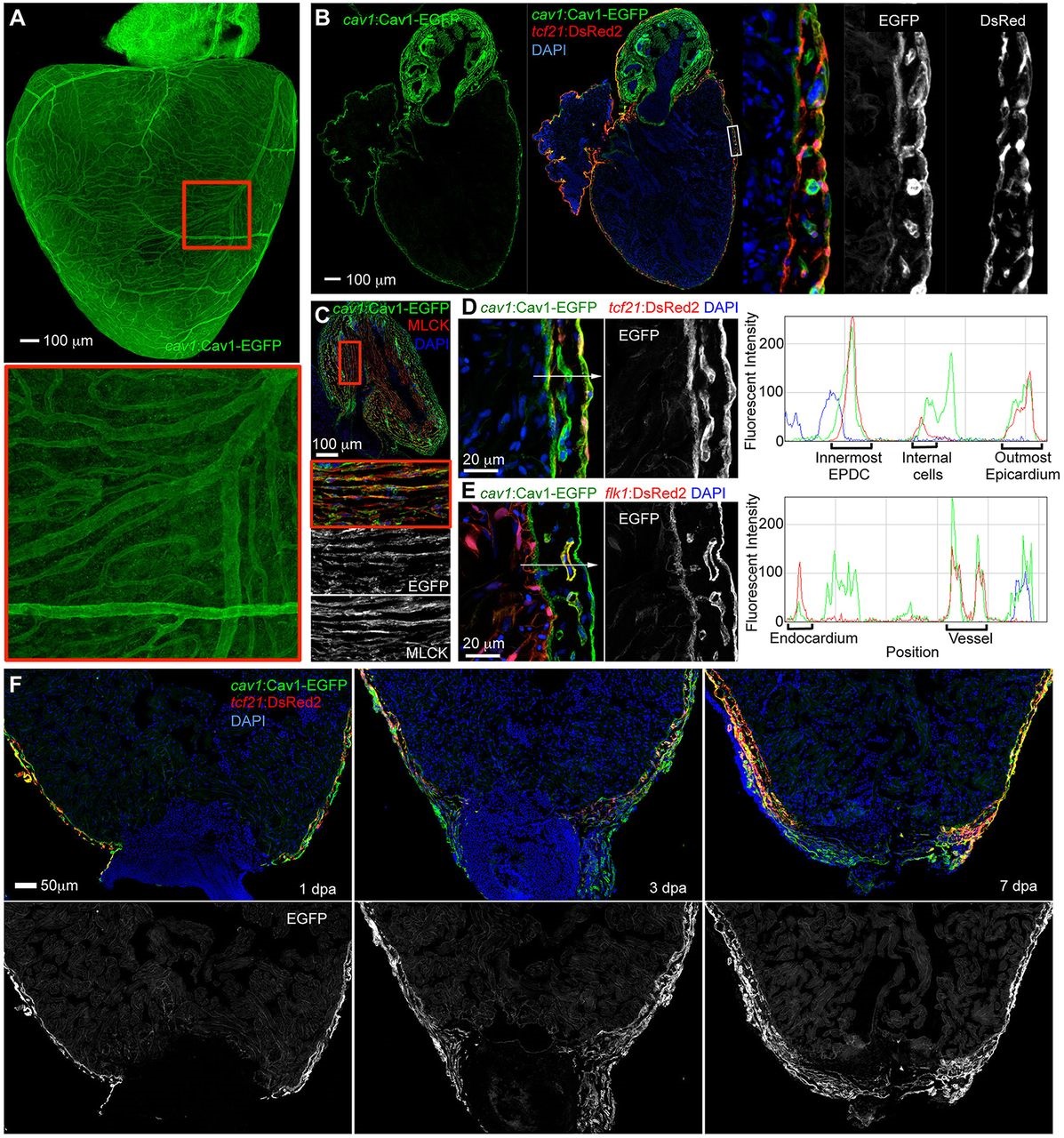
cav1 displays pan-epicardial expression. (A) Cav1-EGFP reporter expression in a whole-mount adult heart, showing labeling of the entire cardiac surface, including coronary vessels. The boxed region is magnified beneath. (B) Tissue sections indicating that the Cav1-EGFP reporter is expressed in epicardial cells, colocalizing with tcf21:DsRed2 signals. Insets show a higher magnification view of the framed region. (C) Section of outflow tract of cav1:cav1-EGFP reporter fish, stained for the smooth muscle marker Myosin light chain kinase (MLCK) (red), indicating smooth muscle expression of the reporter. The boxed region is magnified beneath. (D,E) Ventricular sections showing Cav1-EGFP reporter expression (green) together with tcf21:DsRed2 or flk1:DsRed2 reporter expression, respectively (red). Nuclei are stained with DAPI (blue). Red/blue/green (RGB) plots along the arrowed lines were generated in ImageJ. Green signals colocalized with red signals in the outermost epicardium, internal cells (likely to be perivascular cells), vascular endothelial cells, innermost EPDCs and endocardium. (F) Upon resection of the ventricular apex, cav1-driven Cav1-EGFP expression (green) is prominent in epicardial cells near the injury site at 1 and 3dpa, and covering the wound at 7dpa.