
Image
|
Figure Caption
Fig. S1
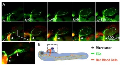
Penetration of endothelial cords into xenografted microtumor on Tg(flk:eGFP::Gata1:dsRED) double transgenic zebrafish.
(A) Real-time observation showing tumor-induced endothelial sprouts (panel t0, arrowhead in magnified image) from the dilated host vessel (arrow in A) on 1dpi are solid and tend to attract each other as they stretching in the microtumor mass. (B) Diagram shows the strategy of establishing the xenograft tumor model by injecting 500-1000 tumor cells besides 48hpf zebrafish common cardinal vein (CCV).
Acknowledgments
This image is the copyrighted work of the attributed author or publisher, and
ZFIN has permission only to display this image to its users.
Additional permissions should be obtained from the applicable author or publisher of the image.
Full text @ Sci. Rep.