
|
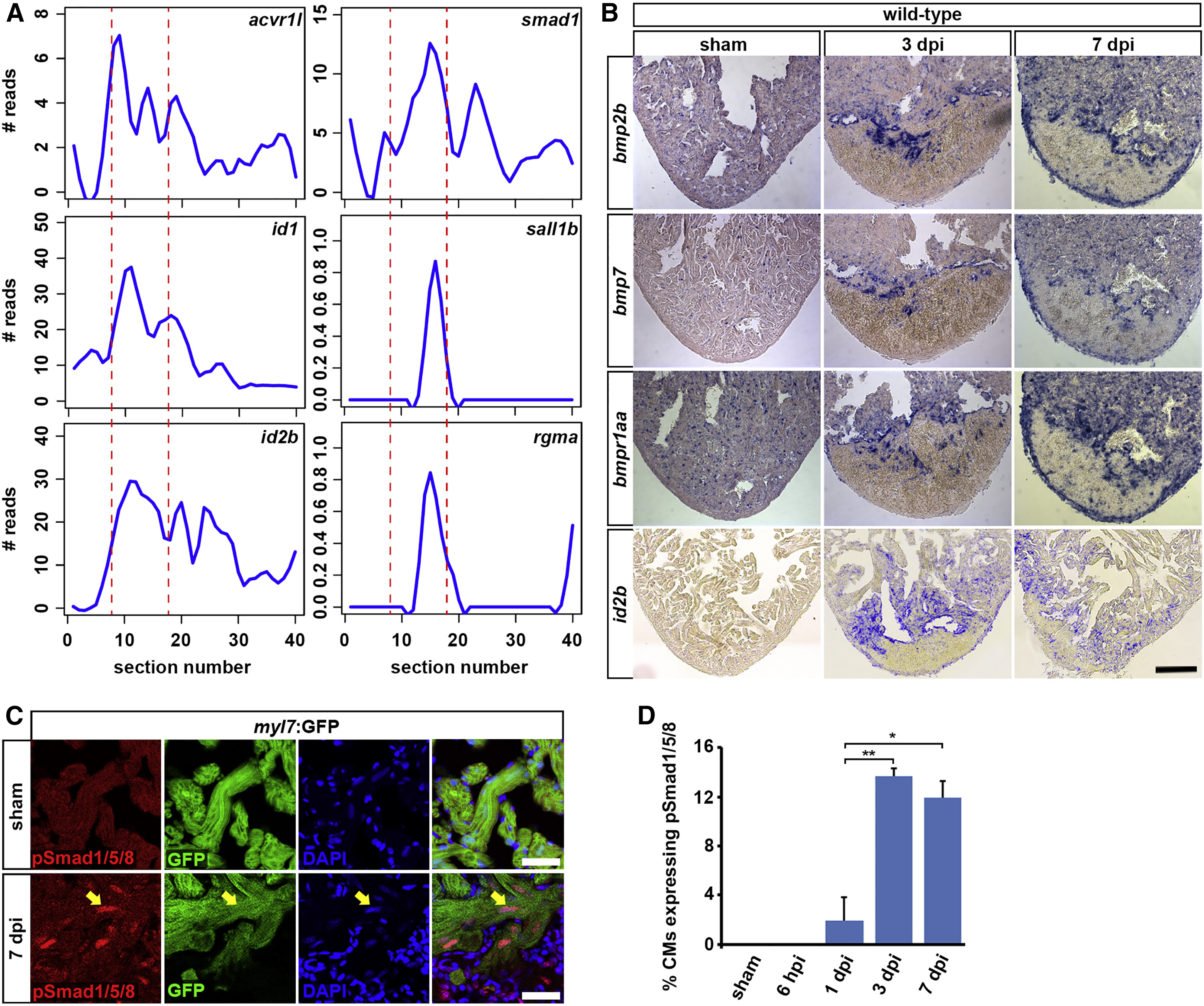
Fig. 4
BMP Signaling Is Activated in Border Zone Cardiomyocytes
(A) Expression traces from tomo-seq data on cryoinjured heart (3 dpi) for BMP signaling-related genes reveal peak expression in the wound border zone (indicated by dashed lines).
(B) In situ hybridizations on sham-operated and 3 and 7 dpi hearts for the BMP ligands bmp2b and bmp7, the BMP receptor bmpr1aa (alk3a), and the BMP target gene id2b. Scale bar, 100 µm.
(C) At 7 dpi, nuclear pSmad1/5/8 expression (arrow) is detected in myl7:GFP-positive cardiomyocytes at the wound border. Scale bar, 25 µm.
(D) Average percentage of cardiomyocytes at the wound border expressing nuclear pSmad1/5/8 at different time points after cryoinjury. Error bars represent SEM. Student’s t test, *p = 0.011, **p = 0.004.
Reprinted from Developmental Cell, 36(1), Wu, C.C., Kruse, F., Vasudevarao, M.D., Junker, J.P., Zebrowski, D.C., Fischer, K., Noël, E.S., Grün, D., Berezikov, E., Engel, F.B., van Oudenaarden, A., Weidinger, G., Bakkers, J., Spatially Resolved Genome-wide Transcriptional Profiling Identifies BMP Signaling as Essential Regulator of Zebrafish Cardiomyocyte Regeneration, 36-49, Copyright (2016) with permission from Elsevier. Full text @ Dev. Cell