
|
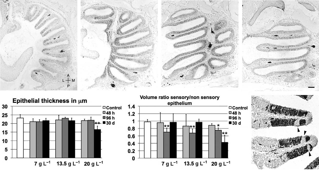
Fig. 1
Morphological analysis of olfactory epithelium before and after treatment. (A) Semi-serial horizontal Hu-positive sections (separated by 100 µm) of untreated olfactory rosette at progressively more ventral planes. A, anterior; L, lateral; M, medial; P, posterior; scale bar: 50 µm. (B) Variations in epithelial thickness across treatments. (C) Comparison between volumes of sensory and non-sensory regions in olfactory mucosa; significant differences are indicated by asterisks: *P < 0.05; **P < 0.01. (D) Calretinin-positive lamellae in zebrafish treated with 7 g L-1 for 96 h; arrowheads: non-sensory areas inserted in sensory epithelium; scale bar: 20 µm.