
Image
|
Figure Caption
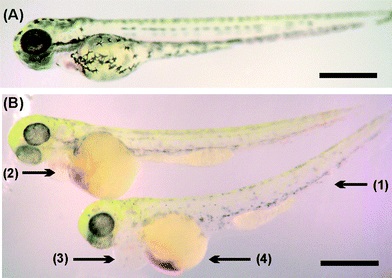
Fig. 4
Morphological changes during incubation with 3D printed materials of 72 hpf zebrafish: a) stereomicrograph of hatched zebrafish embryo control showing normal morphology development. b) Zebrafish after 48 hours of incubation with washed VisiJet Crystal samples. These fish are grossly normal, however they show signs of developmental delay as well as hypopigmentation (1), heart edema (2), bloody pooling (3) and reduced yolk extensions (4). Scale bars are 500 µm.
Acknowledgments
This image is the copyrighted work of the attributed author or publisher, and
ZFIN has permission only to display this image to its users.
Additional permissions should be obtained from the applicable author or publisher of the image.
Full text @ Lab Chip