
Fig. S1
- ID
- ZDB-IMAGE-160212-7
- Publication
- Abu-Siniyeh et al., 2016 - The aPKC/Par3/Par6 polarity complex and membrane order are functionally inter-dependent in epithelia during vertebrate organogenesis
- All Figures
- Figures for Abu-Siniyeh et al., 2016
|
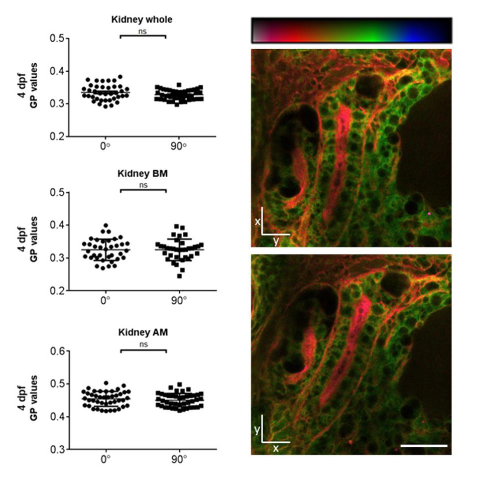
Fig. S1
GP images of zebrafish kidney at different orientations. To address the photoselectivity of the Laurdan dye, zebrafish larvae at 4 dpf were stained with Laurdan and fixed in 4% PFA. Intensity images of the kidneys were recorded with a 2-photon microscope, then the sample was turned by 90° and intensity images of the same organ were taken again. For analysis, the images were converted to GP images. GP images were pseudo-colored with red indicating ordered membranes with high GP values and blue indicating fluid membranes with low GP values, as per color scale. Here GP values of the whole organ, the apical membranes and the basolateral membranes were compared. ns, not significant. Scale bar = 20 µm.