
Fig. 5
- ID
- ZDB-IMAGE-160210-28
- Publication
- Chiavacci et al., 2015 - MicroRNA 19a replacement partially rescues fin and cardiac defects in zebrafish model of Holt Oram syndrome
- All Figures
- Figures for Chiavacci et al., 2015
|
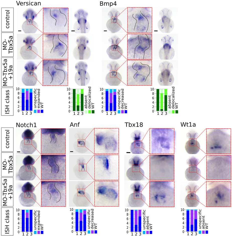
Fig. 5
miR-19a replacement partially rescues cardiac marker expression in Tbx5 morphants.
WT embryos were co-injected with 1.5 ng of MO-Tbx5a and 0.25 ng of miR-Ct (MO-Tbx5a) or 0.25 ng of miR-19a mimics (MO-Tbx5a + miR-19a). WT embryos co-injected with 1.5 ng of MO-Ct and 0.25 ng of miR-CT were used as control. At 48 hpf, 15–20 embryos from each thesis were fixed for ISH analysis. A small number of embryos were also grown up to 72 hpf and screened by optical microscopy to verify that the percentages of embryos with cardiac defects and fin absence were the expected. Whole mount ISH. Ventral view of 48hpf embryos is shown. For versicana and bmp4 probes, dorsal view is also shown (right side) to highlight the fin bud (only bmp4) and fin apical fold signals. Black scale bars:100 µm. At bottom of the figure, quantifications of different hybridization signals were reported. 1 = MO-Ct + miR-Ct; 2 = MO-Tbx5 + miR-Ct and 3 = MO-Tbx5 + miR-19a.