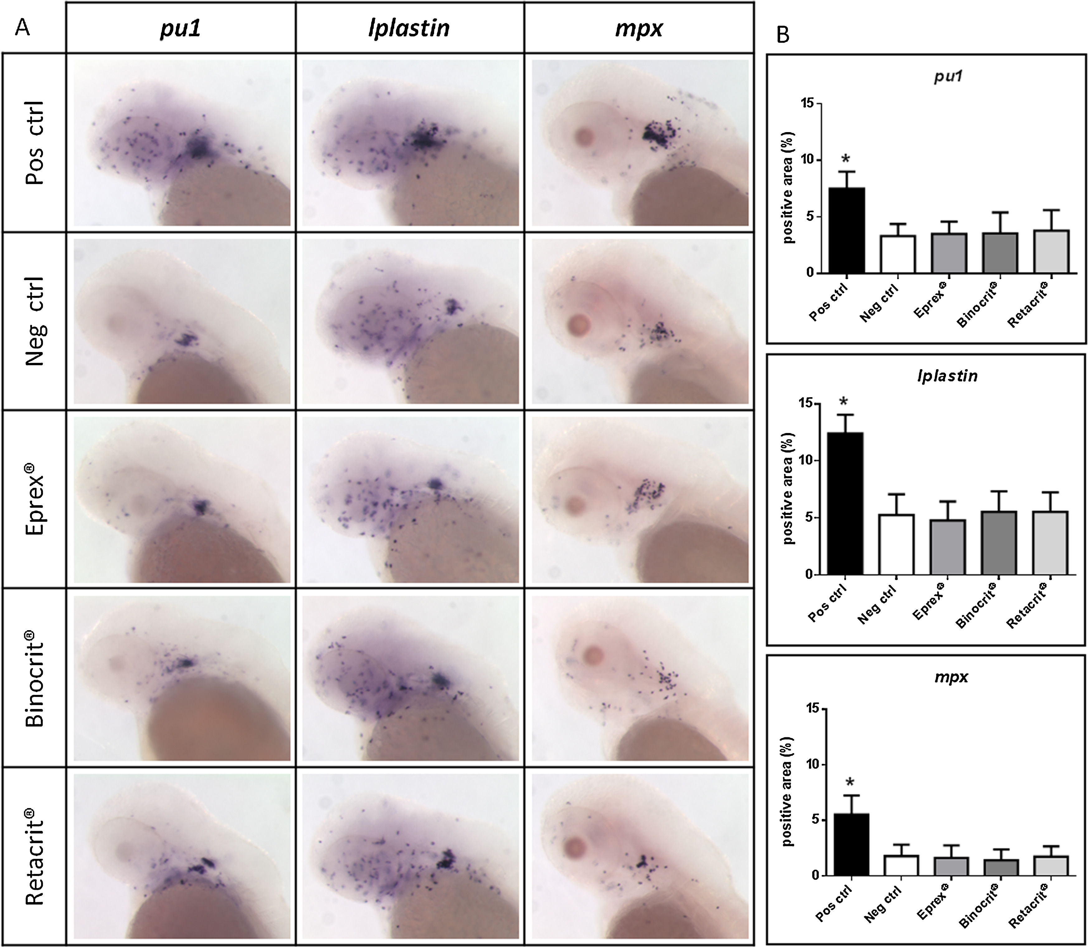
Fig. 6
In situ hybridization with pu1, lplastin and mpx probes to detect leukocytes precursors, monocytes/macrophages and granulocytes neutrophils, respectively. (A) Representative images of 72 hpf embryos injected into the otic capsule with Eprex®, Binocrit® or Retacrit® and positive or negative control (Pos ctrl and Neg ctrl, respectively). Lateral views, anterior to the left, 11.5X magnification; (B) quantification of pu1, lplastin and mpx positive area, measured with ImageJ 1.45 s image analysis software on a mean of 40 embryos for each experimental point. Asterisk indicates statistically significant increase of the positive area (p < 0.05), data are the mean ± S.D. of 3 measurements.

