
Fig. 3
- ID
- ZDB-IMAGE-160210-12
- Genes
- Publication
- Dempsey et al., 2015 - Determination of the source of SHG verniers in zebrafish skeletal muscle
- All Figures
- Figures for Dempsey et al., 2015
|
Fig. 3
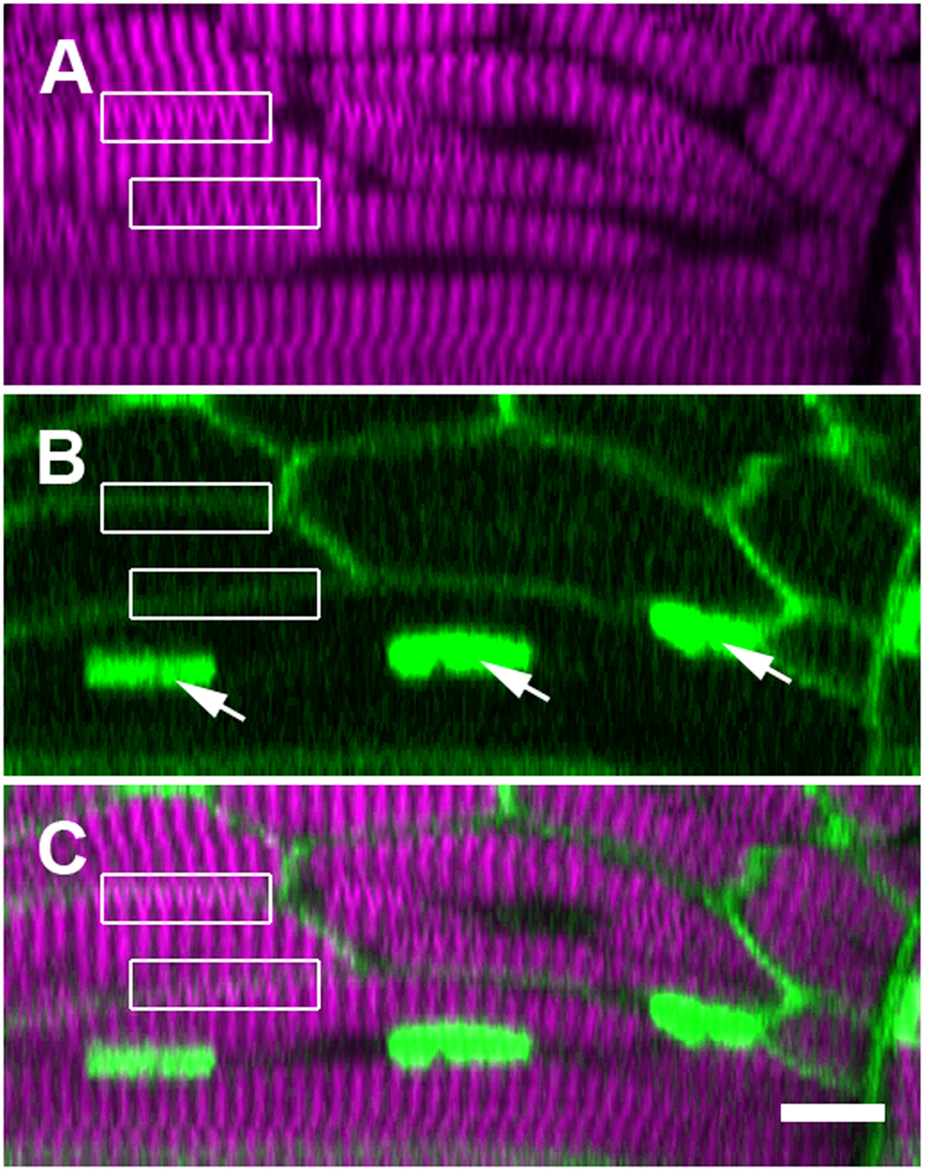
SHG verniers seem to cross intact membrane boundaries, indicating their illusory origin.
These panels display a single ‘xz’ cross section within a trunk somite of a fixed 5 dpf zebrafish larva, where the membranes and nuclei are FP labeled (see Supporting Information, Methods). (A) Verniers (‘Y’-shaped) are visible within much of the thick filament-derived SHG signal (magenta). (B) Fluorescent reporter expression (green) illuminates the plasma membranes (diffuse borders) of the several visible muscle fibers as well as the nuclei within one particular cell (arrows). (C) The merged image of fluorescence and SHG signal shows that many SHG verniers seem to cross the physical membrane boundaries of the muscle cells (boxed regions, compare to the same boxed regions in panel A,B). Sarcomeric components should not connect across adjacent cell boundaries under normal physiological circumstances, especially in an unperturbed WT larva. Scale bar: 10 µm.