
Fig. 1
- ID
- ZDB-IMAGE-160208-34
- Publication
- Hen et al., 2015 - Venous-derived angioblasts generate organ-specific vessels during embryonic development
- All Figures
- Figures for Hen et al., 2015
|
Fig. 1
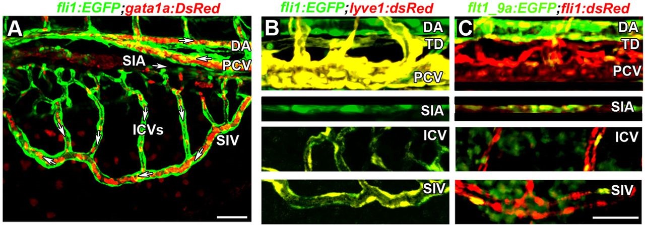
Arterial-venous identity of the subintestinal vessels. (A) Directionality of blood flow in the subintestinal plexus, as determined in Tg(fli1:EGFP;gata1a:dsRed) embryo at 3.5dpf. White arrows indicate the direction of flow in the DA, PCV, SIA, SIV and ICVs. (B) Colocalization (yellow) of fli1:EGFP (green) and lyve1:dsRed fluorescence in venous ECs of the PCV, ICVs, TD and SIV of 4dpf Tg(fli1:EGFP;lyve1:dsRed) double transgenic embryos. The DA and SIA show no lyve1:dsRed expression. (C) Colocalization (yellow) of fli1:dsRed and flt1_9a (green) in arterial ECs of the DA and SIA of 4dpf Tg(flt1_9a_cfos:GFP;fli1:dsRed) double transgenic embryos. DA, dorsal aorta; ICVs, interconnecting vessels; PCV, posterior cardinal vein; SIA, supraintestinal artery; SIV, subintestinal vein; TD, thoracic duct. Scale bars: 50µm.