
|
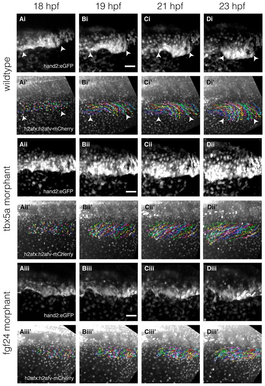
Fig. 2
Time-lapse and cell tracking of the fin-field LPM during pectoral fin bud initiation in wild-type embryos and Tbx5a and Fgf24 morphants. (Ai-Diii′) Time-lapse of double transgenic Et(hand2:eGFP)ch2; Tg(h2afx:h2afv-mCherry)mw3 embryos with backgrounds of wild-type embryos (Ai-Di2), Tbx5a morphant (Aii- Dii′) and Fgf24 morphant (Aiii-Diii′). (Ai-Di,Aii-Dii,Aiii-Diii) Time-lapse stills showing convergence over time of eGFP-expressing fin-field cells. (Ai′-Di′,Aii′-Dii′,Aiii′-Diii′) Corresponding stills of mCherry-labeled nuclei overlaid with cell trajectories indexed by random colors. Dorsal view, anterior left. Scale bars: 50µm. Arrowheads in Ai′-Di′ highlight the extent of the fin-field.